
🎉 Main components of a light microscope. Parts of a microscope with functions and labeled
Microscope Diagram Labeled First and foremost, we have a labeled microscope diagram, available in both black and white and color. Useful as a study guide for learning the anatomy of a microscope. There are six printables available.
Ag Biology Unit 2
Having been constructed in the 16th Century, microscopes have revolutionized science with their ability to magnify small objects such as microbial cells, producing images with definitive structures that are identifiable and characterizable. Derived from Greek words "mikrós" meaning "small" and "skópéō" meaning "look at". Table of Contents
31 Label Diagram Of Microscope Label Design Ideas 2020
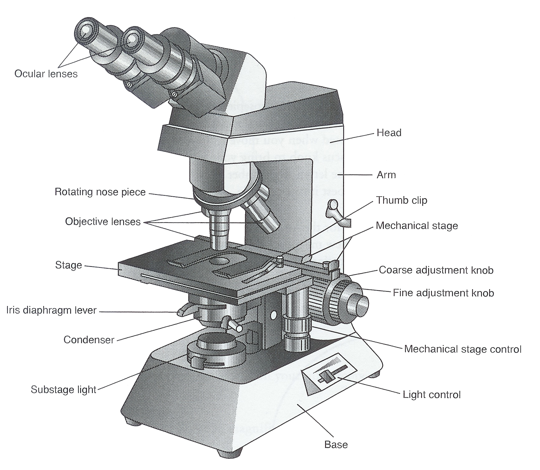
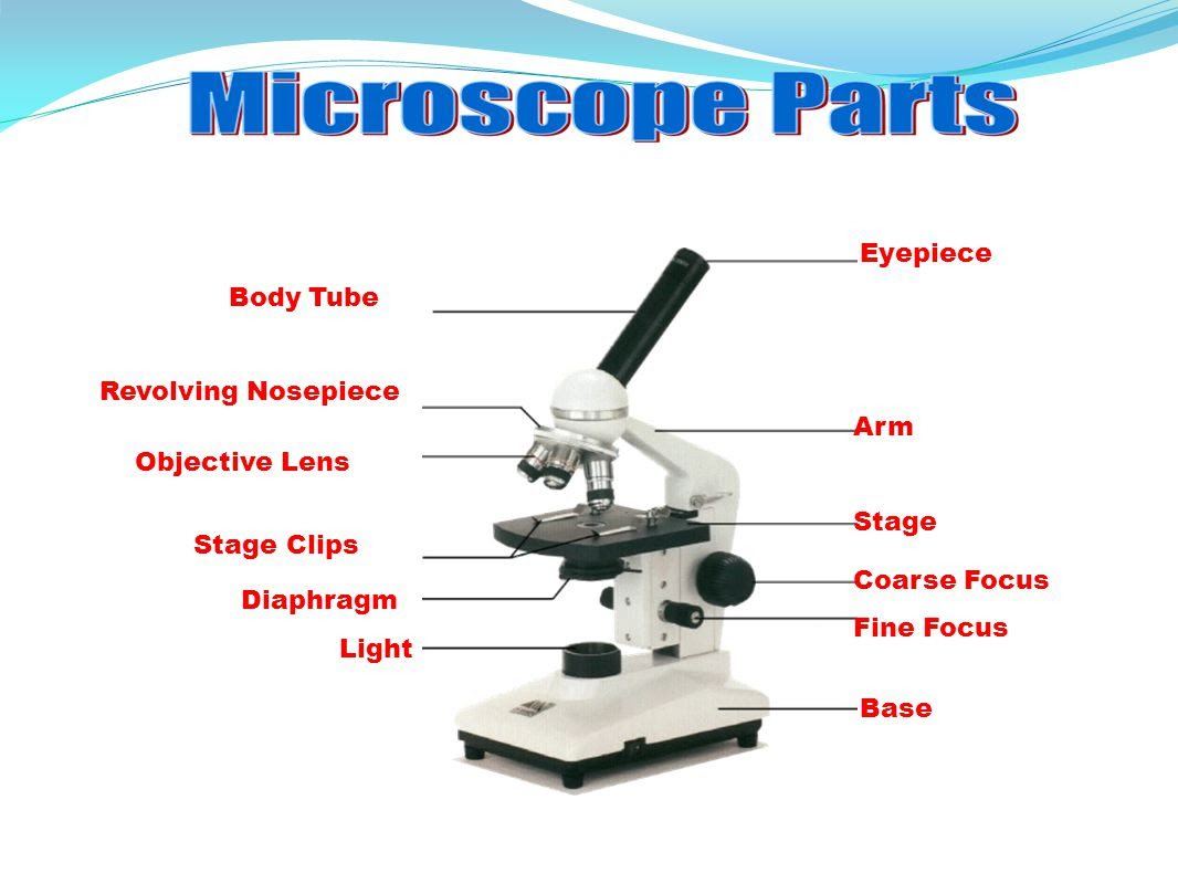
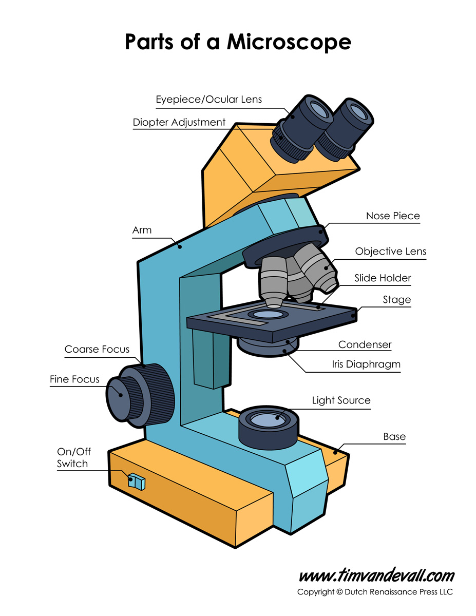
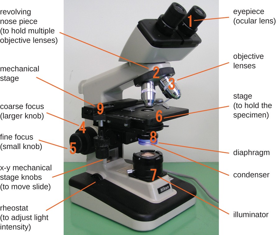
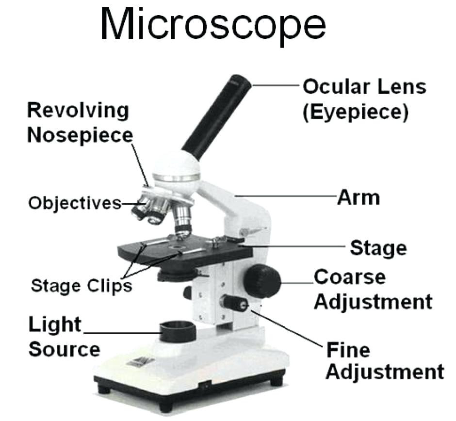
1. Eyepiece or Ocular Lens Eyepiece lens magnifies the image of the specimen. This part is also known as ocular. Most school microscopes have an eyepiece with 10X magnification. 2. Eyepiece Tube or Body Tube The tube hold the eyepiece. 3. Nosepiece Nosepiece holds the objective lenses and is sometimes called a revolving turret.
Monday September 25 Parts of a Compound Light Microscope
Mirror. The lower end of the arm or the pillar has a mirror fastened to it. On one side is a regular mirror, and on the other is a concave mirror. It is used to reflect light into the microscope for a sharper view of the specimen. A compound microscope primarily makes use of concave mirrors. Plane mirrors are occasionally also used.
23 Label And Color The Parts Of Both Microscopes Labels 2021
Blank microscope to label parts. This page titled 1.5: Microscopy is shared under a CC BY 4.0 license and was authored, remixed, and/or curated by Orange County Biotechnology Education Collaborative ( ASCCC Open Educational Resources Initiative ) .
Microscope Labeled Diagram Micropedia Gambaran
A microscope is an instrument that magnifies objects otherwise too small to be seen, producing an image in which the object appears larger. Most photographs of cells are taken using a microscope, and these pictures can also be called micrographs. From the definition above, it might sound like a microscope is just a kind of magnifying glass.

How to Use a Microscope (Properly) Step by Step New York Microscope Company
Open combination drawer and take out the microscope. 3. Label all the parts of the microscope with the provided post-its using the image below or the laboratory manual. Note. The image below does not match your microscope perfectly, you will be responsible for knowing the parts of your microscope on the lab practical.
Dissecting Microscope Labeled Diagram Micropedia Images and Photos finder
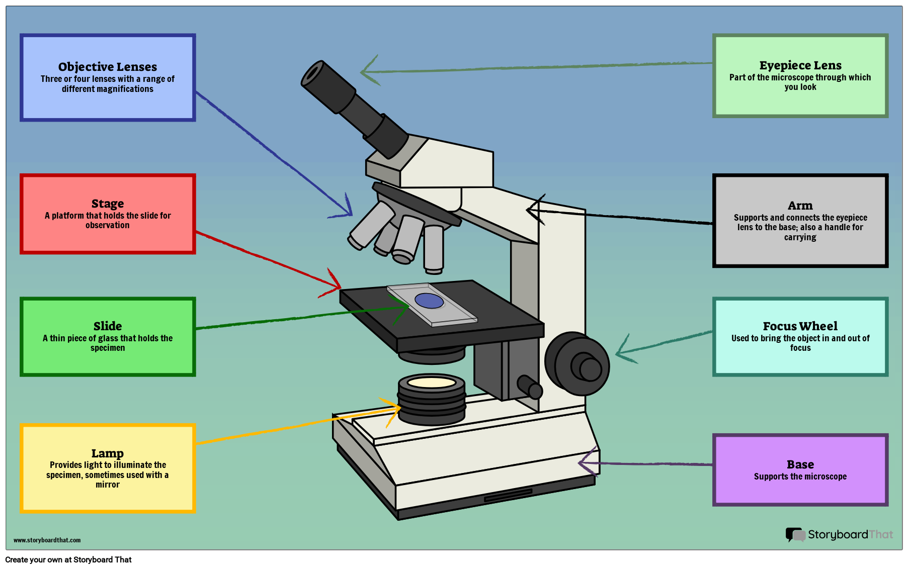
It also allows the specimen to be labeled, transported, and stored without damage. Stage: The flat platform where the slide is placed. Stage clips: Metal clips that hold the slide in place. Stage height adjustment (Stage Control): These knobs move the stage left and right or up and down.
Parts of a Compound Microscope — Learning in Hand with Tony Vincent
August 2017. Clip art purchased from StockPhoto and used with permission. Parts of a Microscope. Label the parts of the microscope. You can use the word bank below to fill in the blanks or cut and paste the words at the bottom. Created by Jolanthe @ HomeschoolCreations.net. Parts of a Microscope.

Microscope, Microscope Parts, Labeled Diagram, and Functions
There are 1000 millimeters (mm) in one meter. 1 mm = 10 -3 meter. There are 1000 micrometers (microns, or µm) in one millimeter. 1 µm = 10 -6 meter. There are 1000 nanometers in one micrometer. 1 nm = 10 -9 meter. Figure 1: Resolving Power of Microscopes. The microscope is one of the microbiologist's greatest tools.
36+ Label Each Part Of A Microscope Gif Diagram Printabel
Parts of the Microscope with Labeling (also Free Printouts) A microscope is one of the invaluable tools in the laboratory setting. It is used to observe things that cannot be seen by the naked eye. Table of Contents 1. Eyepiece 2. Body tube/Head 3. Turret/Nose piece 4. Objective lenses 5. Knobs (fine and coarse) 6. Stage and stage clips 7. Aperture

Label the Microscope Diagram Download Scientific Diagram
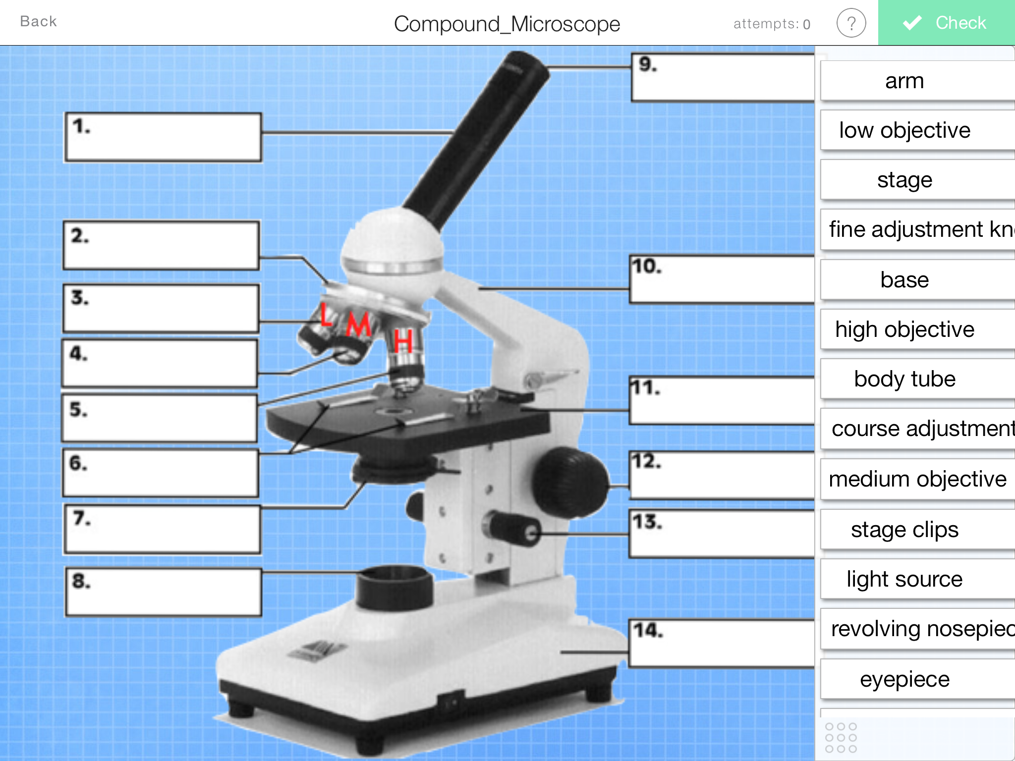
Label the microscope Interactive Add to collection Use this interactive to identify and label the main parts of a microscope. Drag and drop the text labels onto the microscope diagram. base eye piece lens diaphragm or iris fine focus adjustment high-power objective coarse focus adjustment light source stage Download Exercise Tweet

Simple Microscope Diagram (Parts labelled), Principle, Formula and Uses
This simple worksheet pairs with a lesson on the light microscope, where beginning biology students learn the parts of the light microscope and the steps needed to focus a slide under high power.. The labeling worksheet could be used as a quiz or as part of direct instruction. Students label the microscope as you go over what each part is used for.

Parts of a Compound Microscope Labeled (with diagrams) Medical Pictures and Images (2023
No matter what you love, you'll find it here. Search Microscope Label and more. Looking for Microscope Label? We have almost everything on eBay.
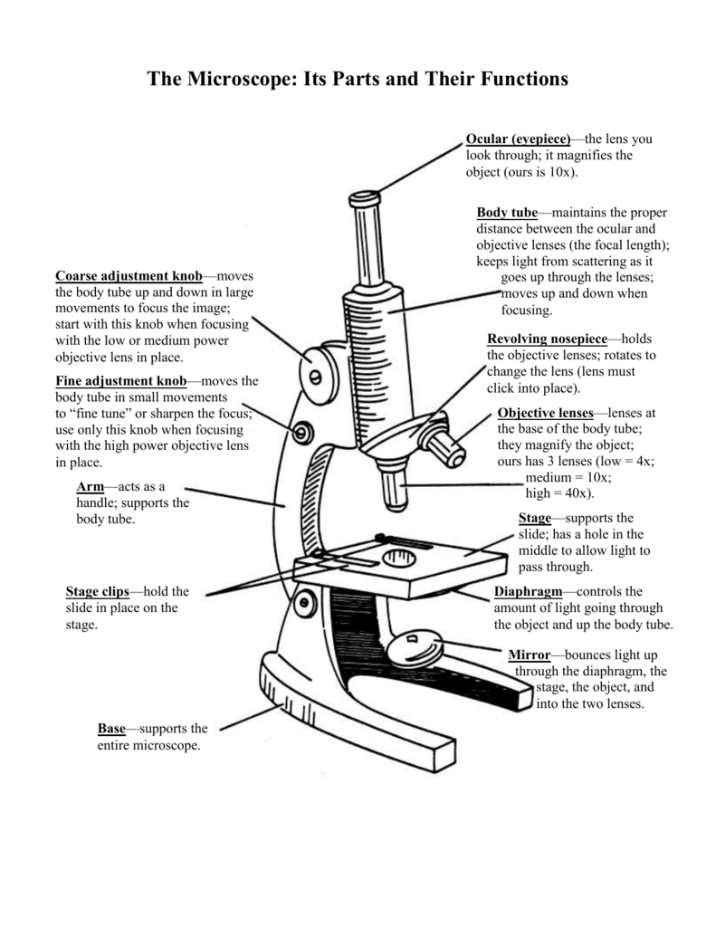
The Microscope Its Parts and Their Functions
This activity has been designed for use in homes and schools. Each microscope layout (both blank and the version with answers) are available as PDF downloads. You can view a more in-depth review of each part of the microscope here. Download the Label the Parts of the Microscope PDF printable version here.

Parts of a microscope with functions and labeled diagram
Laboratory Labels - Use Our Truly Streamlined Ordering Process. Shop Now! Our Lab Labels are available For Thermal Transfer, Laser & Inkjet Printers.
