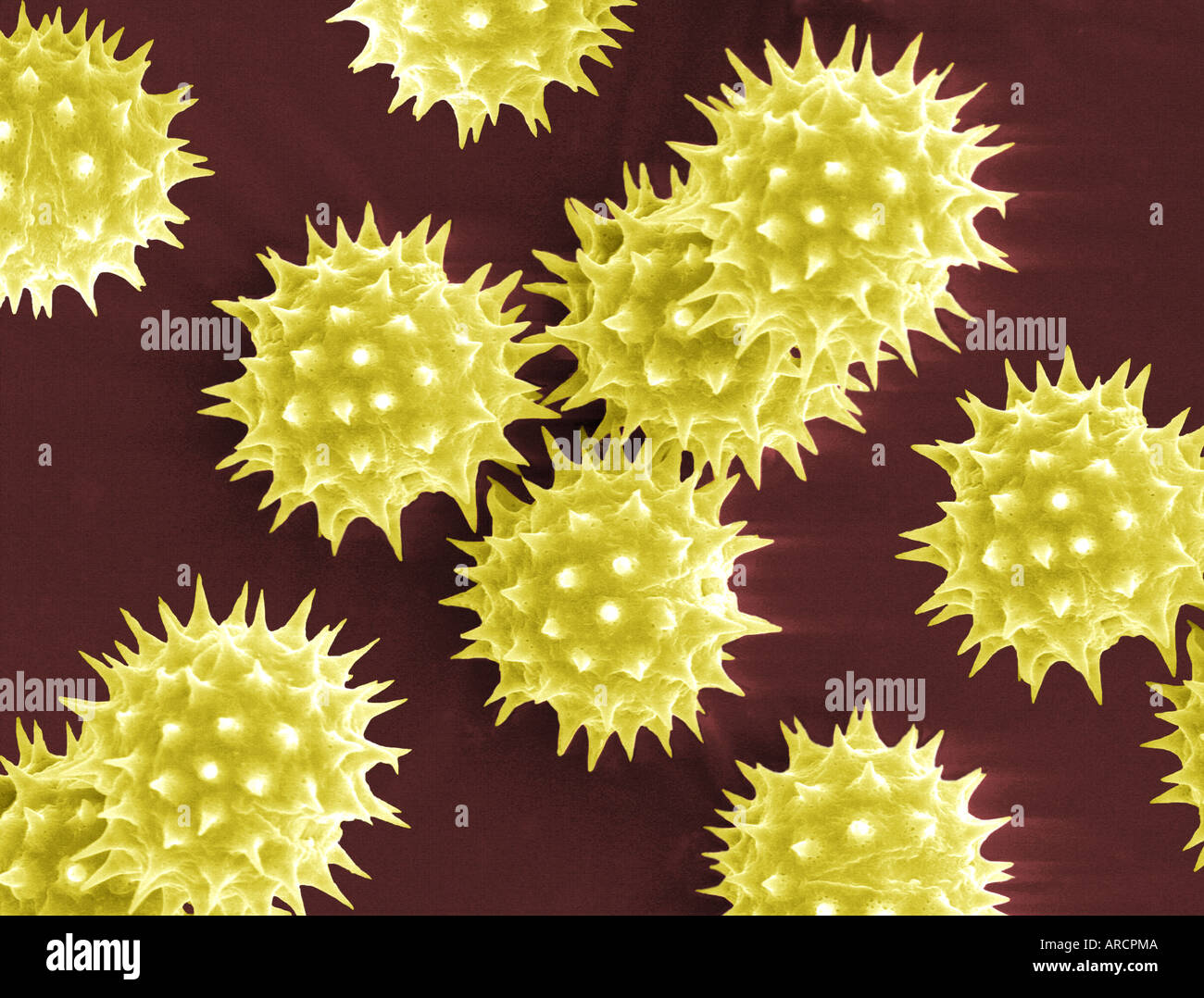
Scanning electron microscope image of pollen grains from Helianthus Stock Photo 9150665 Alamy
Therefore, in this research, light microscopy and scanning electron microscopy were used to observe seven morphological traits of pollens from 22 common vetch accessions, and residual maximum likelihood and pattern analysis was conducted.

Pollen morphology observed under scanning electron microscopy. Upper... Download Scientific
Hitherto such studies have used optical or transmission electron microscopy but here a recently devised preparative technique has enabled pollen development in Cosmos bipinnatus to be studied using the scanning electron microscope. The technique involves freeze-fracturing of osmium fixed, cryoprotected anthers, maceration in dilute osmium.

A variety of pollens. Microscopic, Electron microscope, Microscopic images
This study employs scanning electron microscopy (SEM) to delve into the intricate pollen morphology of Cucurbitaceae (Gourd Family) species, unraveling the nuanced details of their structural features. Concurrently, the research investigates the antimicrobial potentials encoded within these pollen.
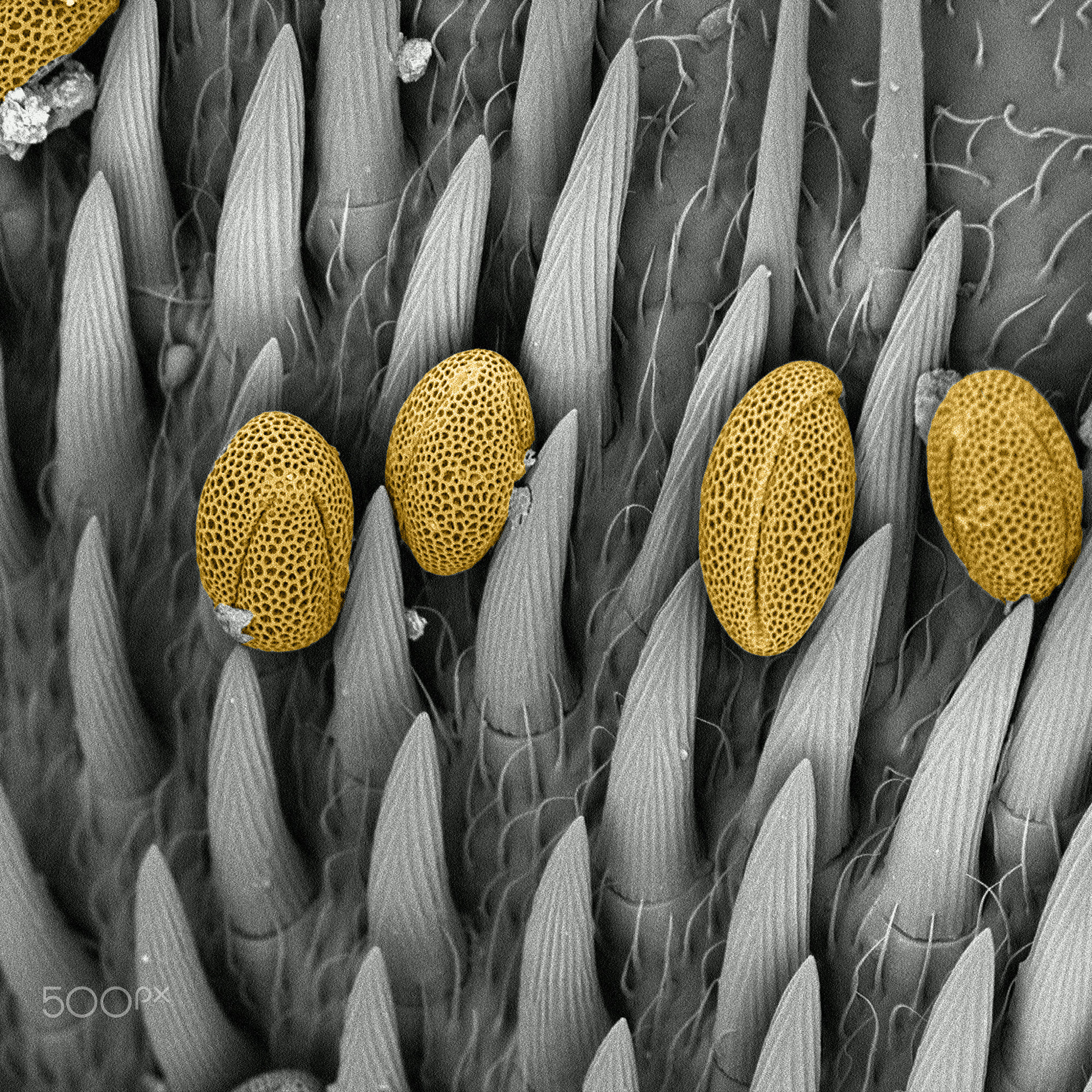
pollen on bumblebee scanning electron microscope by strucTEMART microscopic ART Photo
This Scanning Electron Microscopic image reveals pollen grains from a variety of common plants: sunflower (Helianthus annuus), morning glory (Ipomoea purpurea ), prairie hollyhock (Sidalcea malviflora), oriental lily (Lilium auratum ), evening primrose (Oenothera fruticosa), and castor bean (Ricinus communis). Download

POLLEN under electron microscope Microscopic photography, Microscopic images, Electron
SMOOTH OVER Pollen grains from flowering plants can be relatively smooth (one shown in this scanning electron microscope image at left). Computer simulations of pollen formation show that halting.
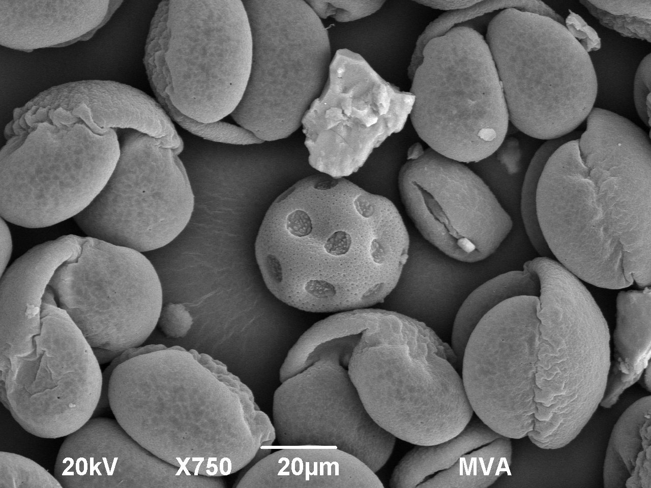
Pollen Grains Under the Microscope MVA Scientific Consultants
The electron-micrographs were made using a Quanta 250 microscope (FEI Company) and JEOL 6390LV microscope. Descriptions follow Punt et al. (2007) , and the produced slides were deposited at the pollen library of Plant Micromorphology Laboratory (LAMIV), of the State University of Feira de Santana.

Grains of pollen as seen by an electron microscope Boing Boing
Here, we describe methods of transmission electron microscopy (TEM) based on conventional chemical fixation and high-pressure freezing (HPF) and freeze-substitution (FS) to examine the ultrastructure of Arabidopsis pollen grains and pollen tubes.

Pollen under an electron microscope pollen microscope Flickr
Free Shipping Available. Buy An Electron Microscope on ebay. Money Back Guarantee!
GMS Scanning Electron Microscope Still Image of Pollen Particles
A scanning electrode microscope ( SEM) is a type of electron microscope that produces images of a sample by scanning the surface with a focused beam of electrons. The electrons interact with atoms in the sample, producing various signals that contain information about the surface topography and composition of the sample.
Electron microscope picture of pollen WQHD_Wallpaper
In this work, the suitability of three microscopic techniques for automatic analysis of pollen grains was studied. 2D and 3D morphological characteristics, textural and colour features, and extended depth of focus characteristics were used for the pollen discrimination.

nature calling coated in pollen
The outermost layer ("exine") is made up of sporopollenin, which is a strong, crosslinked biopolymer 7, while the inner layer ("intine") is composed of elastic, load-bearing cellulose/hemicellulose.

Smithsonian Insider Research collection of pollen grains given to Smithsonian Tropical
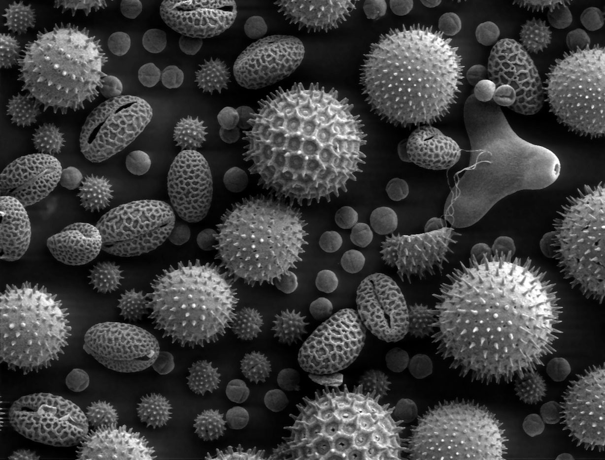
False-colored scanning electron micrographs show the diverse ornamentation patterns on the surfaces of pollen from different species.

Nature Prefers Asymmetrical Pollen Grains, Study Finds News
Microscopy Research and Technique (MRT) is an international, advanced microscopy journal covering the fields of biological, clinical, chemical, & materials sciences.

POLLEN grains under an electron microscope. Photo Courtesy of Dartmouth Electron Microscope
soil analysis. Palynologists rely on light microscopy (LM) to identify and interpret the pollen spectrum of a particular sample. Scanning electron microscopy (SEM) is not normally used for counting and identifying pollen grains. Instead, SEM is mainly used for morphological comparisons and taxonomy where the increased resolution of SEM makes.

Picturing Pollen COLORS OF NATURE
Pollen grains of Campomanesia pubescens (a-c), Caryocar brasiliense (d-f), Erythroxylum campestre (g-i), Lippia lupulina (j-l), Pyrostegia venusta (m-o), and Xylopia aromatica (p-r), under scanning electron microscope. a, d, g, j, m, p Pollen apertures in polar view, the artificially colored areas indicating the colpi (orange) and.

Pollen under a scanning electron microscope (One Bite at a Time)
Accurate and rapid identification of pollen species under the electron microscope help medical staff in pollen forecast and interrupt the natural course of pollen allergy.
