
The Structure and Functions of an Animal Cell
Animal Cell- Definition, Structure, Parts, Functions, Labeled Diagram. By Go Life Science Posted on December 20, 2022. An animal cell is a type of cell that is characteristic of animals and is present in all multicellular organisms that belong to the animal kingdom. Animal cells are eukaryotic, which means they have a true nucleus that holds.

Ribosomes Job Animal Cell / Nucleolus, Nucleous, Ribosomes, and Vacuole
The animal body has several types of cells. Examples of common animal cell types include skin cells, muscle cells, blood cells, fat cells, nerve cells, sex cells, and stem cells. Skin cells are cells that make up the skin or epithelial tissue. Muscle cells (also called myocytes) are cells that make up muscular tissue.

What is a cell? Facts
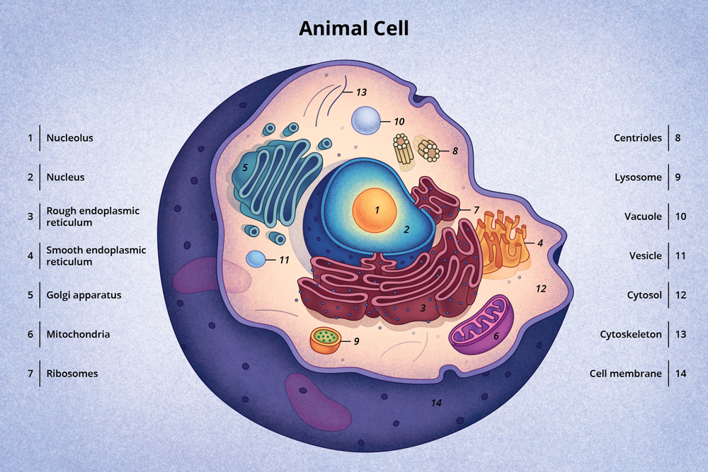
Image 1: Structure of Animal cell (diagram) Image created with the help of biorender Difference between Prokaryotic Cell and Eukaryotic Cell Prokaryotic cells are unicellular that do not contain membrane-bounded organelles. Their nucleic material is dispersed in the cytoplasm.

What Is An Animal Cell? Facts, Pictures & Info For Kids & Students.
Animal cells perform multiple functions essential for the survival and adaptation of organisms. Animal cell is eukaryotic in nature and exhibit DNA within the nucleus. It also contains various cellular structure and organelles like the cytoplasm, Golgi apparatus, ribosomes, mitochondria, etc. Animal cells function together to carry out various.
Animal eukaryotic cell structure
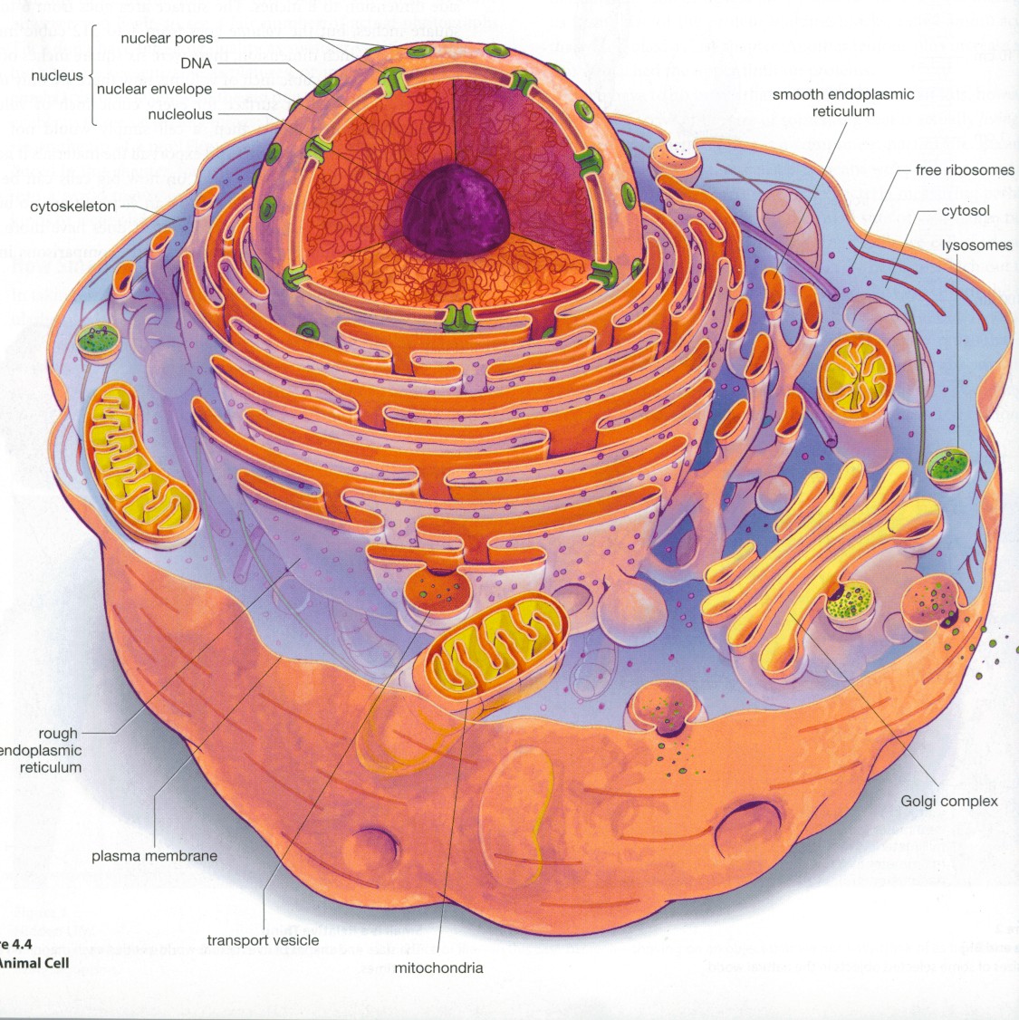
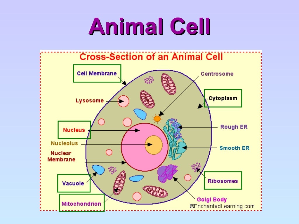
Animal Cell Diagram and Structure (cross section of an animal cell) The cell of the animal is composed of a variety of structural organelles that are enclosed within the plasma membrane. These organelles allow it to function effectively by triggering mechanisms that are beneficial to those who are the hosts (animal). The interaction of all.

Biology 101 Cells Owlcation
Animal cells are the fundamental units of life in protozoa and multicellular animals. Each cell is a wonder in its own right, plus they work together as building blocks for tissues, organs, and organ systems. Animal cells are mostly microscopic, ranging in size from 1 to 100 micrometers.

South Pontotoc Biology Plant and Animal Cell Diagrams
Definition Animal cells are the basic unit of life in organisms of the kingdom Animalia. They are eukaryotic cells, meaning that they have a true nucleus and specialized structures called organelles that carry out different functions.
Discovery and Structure of Cells Biology Visionlearning
Key points: All cells have a cell membrane that separates the inside and the outside of the cell, and controls what goes in and comes out. The cell membrane surrounds a cell's cytoplasm, which is a jelly-like substance containing the cell's parts. Cells contain parts called organelles. Each organelle carries out a specific function in the cell.
Animal Cell Structure Basic / Cell Structure Cells Structure and
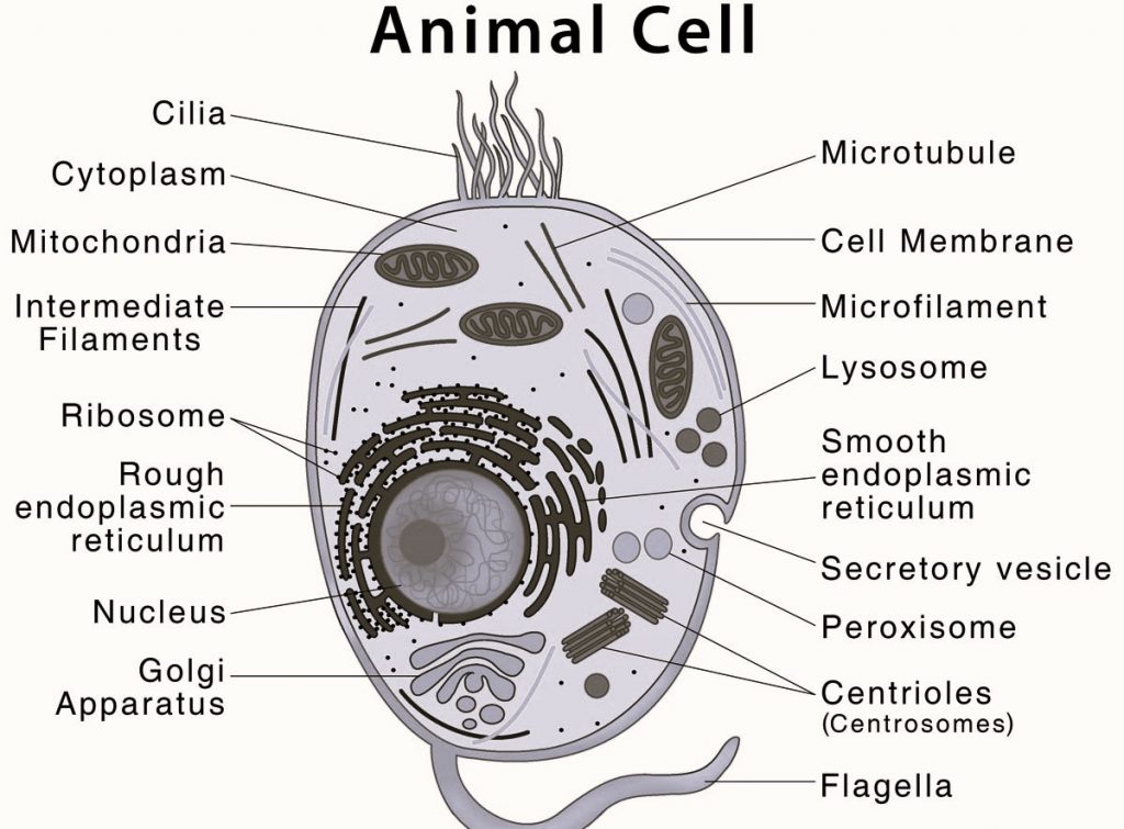
An animal cell is a eukaryotic cell that lacks a cell wall, and it is enclosed by the plasma membrane. The cell organelles are enclosed by the plasma membrane including the cell nucleus. Unlike the animal cell lacking the cell wall, plant cells have a cell wall.
Animal Cell Diagram CBSE Class Notes Online Classnotes123
Animal Cell Diagram. 1) Centrioles. They are paired tube-like organelle composed of a protein called tubulin. Centrioles are about 500nm long and 200nm in width that are found close to the nucleus and helps in cell division.They are also found in cilia and flagella.. Functions

Animal Cell diagram with labels by Russell Kightley Media
Animal cell diagram detailing the various organelles Though this animal cell diagram is not representative of any one particular type of cell, it provides insight into the primary organelles and the intricate internal structure of most animal cells.

animal cell diagram easy Kris Hammett
All animal cells are surrounded by a protective membrane which is called as cell-membrane or plasma membrane. It is also called as cytoplasmic membrane. Plasma membrane is a thin, elastic and semi-permeable membrane. It is mainly composed of 32% lipids, 12% protein, 6% carbohydrates and 20% water. The pores of the membrane allow the passage of.

The animal cell diagram. Vector illustration on white Etsy in 2021
Animal and plant cells share common elements like plasma membranes, cytoskeletons, and mitochondria. However, they differ in certain aspects. For example, plant cells have a cell wall and a central vacuole, while animal cells contain centrosomes.. The lipid bilayer is a more general term that refers to the structure of the membrane itself.
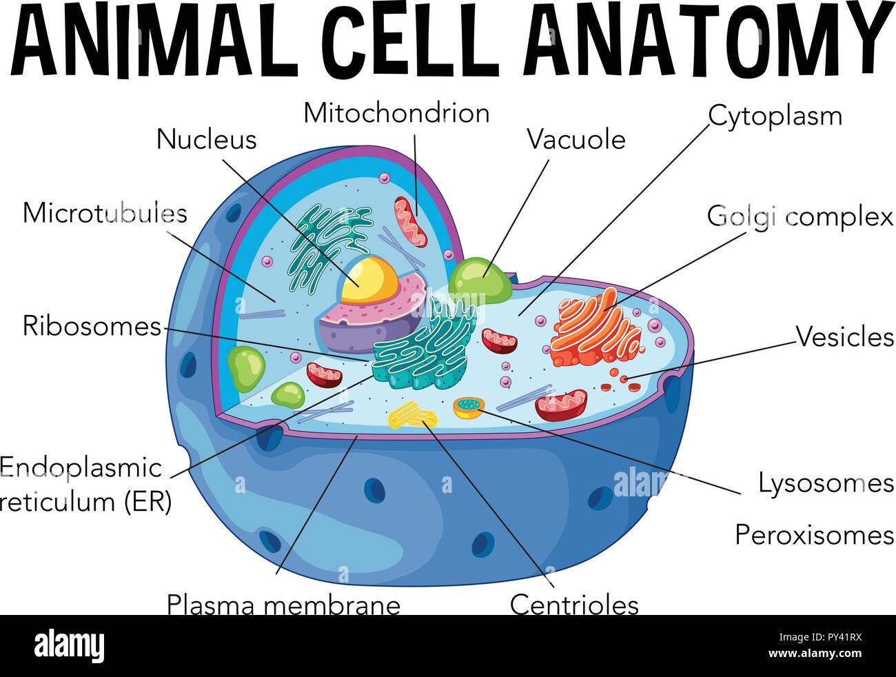
Diagram of animal cell anatomy illustration Stock Vector Image & Art
A Labeled Diagram of the Animal Cell and its Organelles There are two types of cells - Prokaryotic and Eucaryotic. Eukaryotic cells are larger, more complex, and have evolved more recently than prokaryotes. Where, prokaryotes are just bacteria and archaea, eukaryotes are literally everything else.
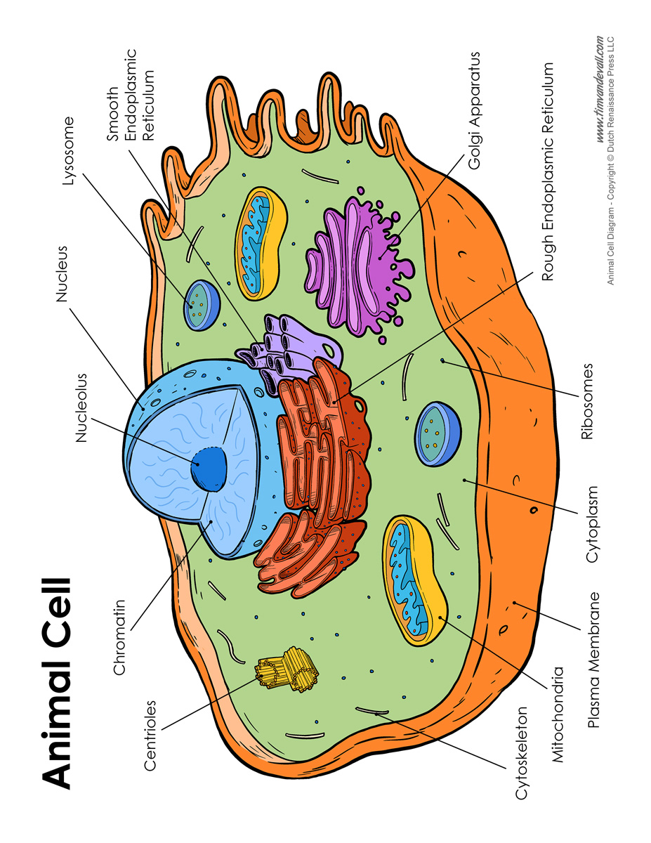
Animal Cell Diagram Labeled Tim van de Vall
Cell Membrane The thin layer of protein and fat that surrounds the cell. The cell membrane is semipermeable, allowing some substances to pass into the cell and blocking others. Centrosome (Microtubule Organizing Center) A small body located near the nucleus - it has a dense center and radiating tubules.

Cells
Looking for a Labeled Diagram of an Animal Cell? You Found It! What's inside an animal cell? Animal Cells are made up of a number of unique parts, including: Cell Membrane Nucleus Nuclear Membrane Centrosome Lysosome Cytoplasm Golgi Apparatus And more! Create Your Own Animal Cell Diagram - Printable Animal Cell Worksheets and More!
