Easy Notes On 【Nasal Cavity】Learn in Just 4 Minutes! Earth's Lab
The cranium (skull) is the skeletal structure of the head that supports the face and protects the brain. It is subdivided into the facial bones and the brain case, or cranial vault ( Figure 7.3 ). The facial bones underlie the facial structures, form the nasal cavity, enclose the eyeballs, and support the teeth of the upper and lower jaws.
Medial wall of the nasal cavity Anatomy and structure Kenhub
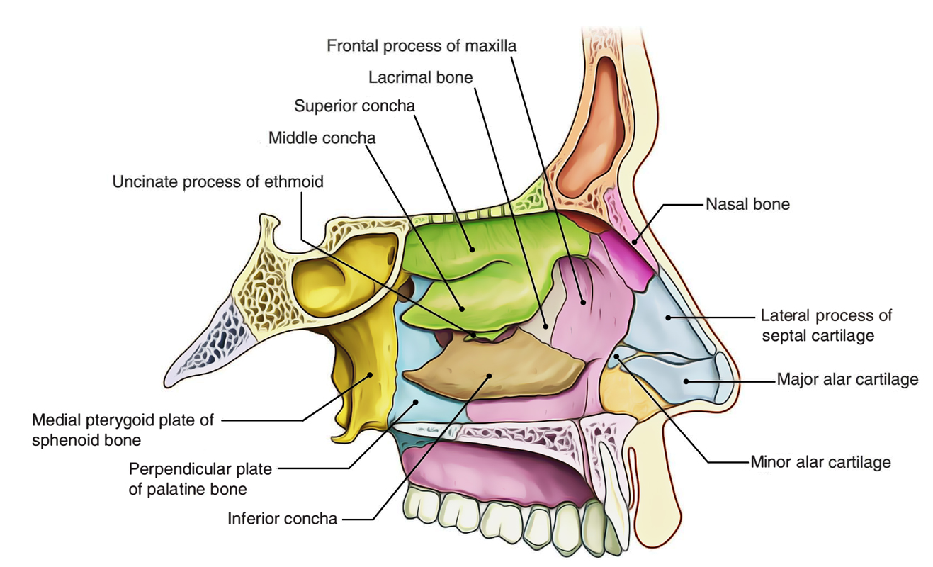
The medial wall, nasal wall, or base of maxillary sinus presents, in the disarticulated bone, a large, irregular aperture, communicating with the nasal cavity. In the articulated skull this aperture is much reduced in size by the following bones: the uncinate process of the ethmoid above, the ethmoidal process of the inferior nasal concha below, the vertical part of the palatine behind, and a.
Nasal Cavity
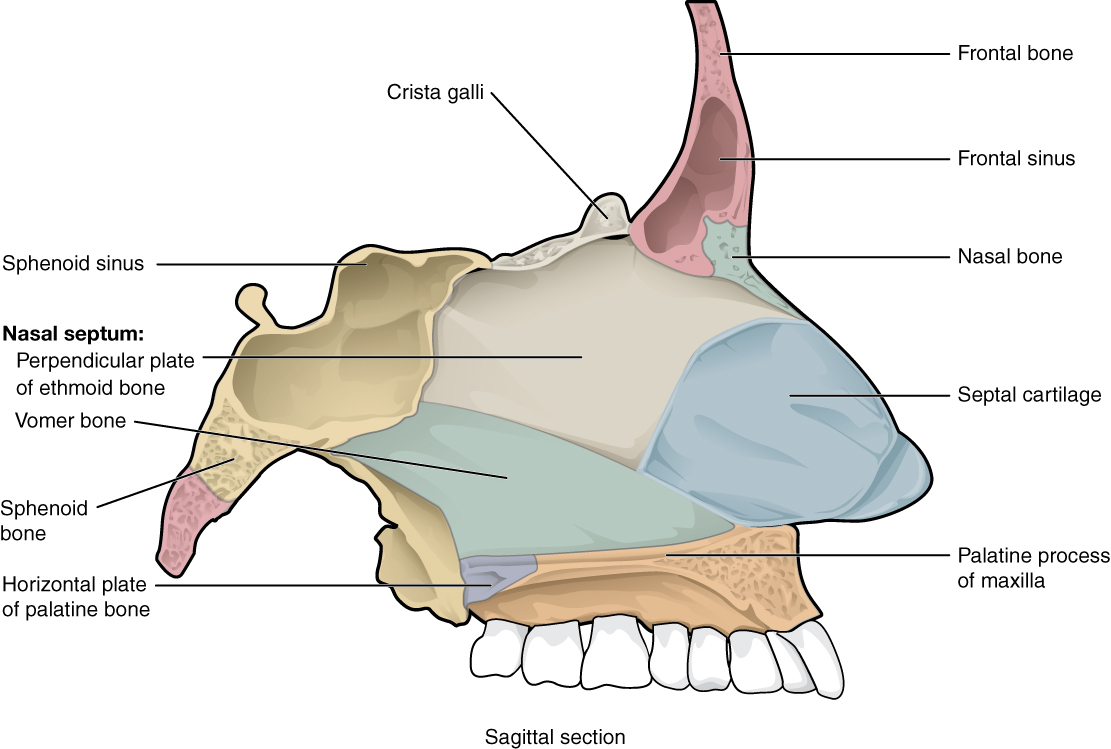
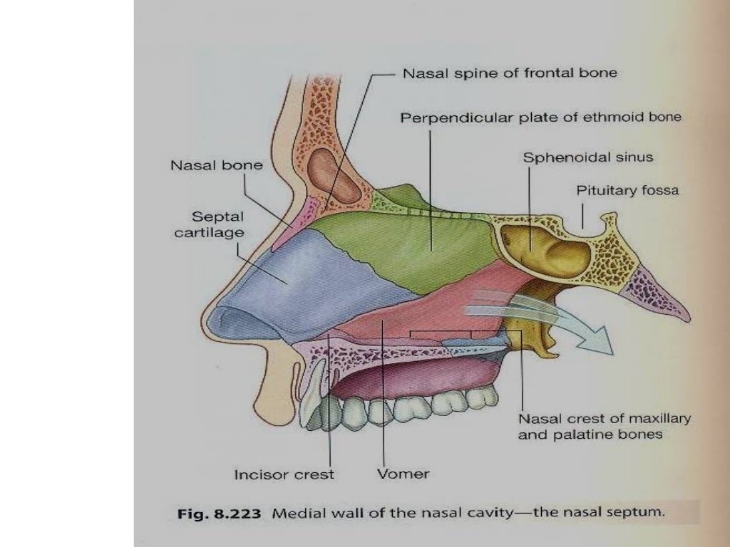
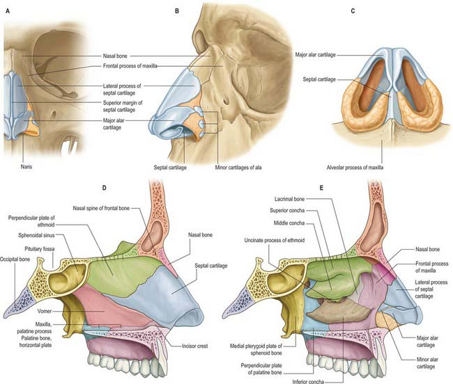
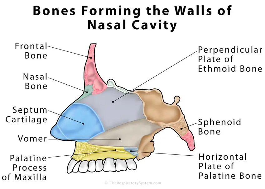
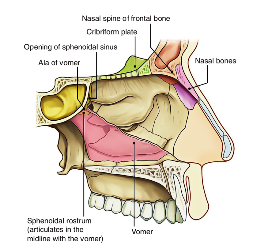
Bones, cartilages and mucosa of the medial wall of the nasal cavity. Nasal septum Septum nasi 1/12 Synonyms: none The nasal septum is the midline vertical partition which separates the nasal cavity into left and right halves, forming the medial wall of each half. It is comprised of bony and cartilaginous parts.
Nariz y Cavidad Nasal Anatomía Concise Medical Knowledge
The medial wall of the nasal cavity is formed by the nasal septum. Anteriorly, it is continuous with the septal cartilage of the external nose. Posteriorly, it Is formed by the bony perpendicular plate of ethmoid (superiorly) and the vomer (inferiorly). Complete Anatomy The world's most advanced 3D anatomy platform Try it for Free

Medial Wall of the Nasal Cavity (preview) Human Anatomy Kenhub YouTube
The lateral wall of the nasal cavity is a region of the nasopharynx essential for humidifying and filtering the air we breathe in nasally. Here we can find a structure called agger nasi. The agger nasi is also referred to as the 'nasoturbinal concha' or 'nasal ridge.'

Medial wall of nasal cavity (nasal septum) anatomy images illustrations anatomy images
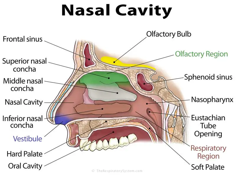
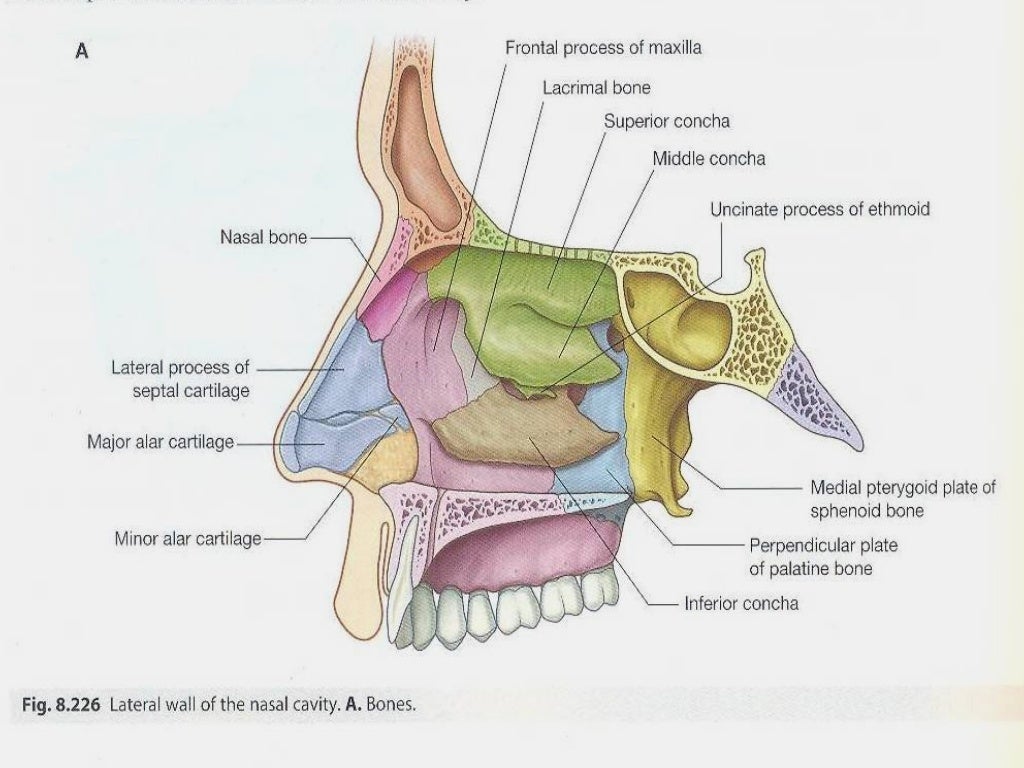
These pathways are called meatuses: Inferior meatus - between the inferior concha and floor of the nasal cavity. Middle meatus - between the inferior and middle concha. Superior meatus - between the middle and superior concha. Spheno-ethmoidal recess - superiorly and posteriorly to the superior concha.

PPT Anatomy of Nose and Paranasal Sinus PowerPoint Presentation ID2646477
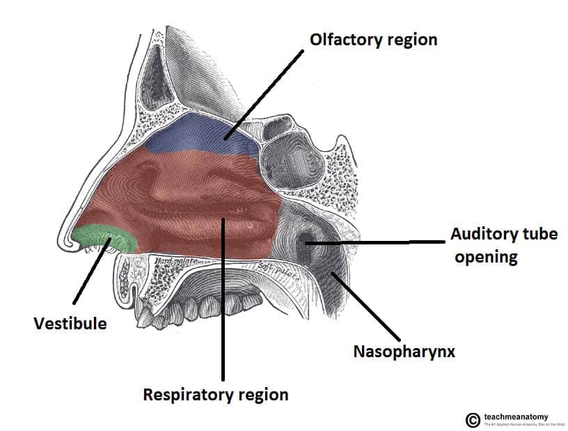
The nasal cavity is lined with a mucous membrane (a lining of tissue) that makes mucus to help keep your nose moist and prevent nose bleeds from a dry nose. There are also little hairs, called cilia, on the inside walls of the nose that filter the air you breathe in to prevent dust and dirt from getting into your lungs.
Nasal Cavity Anatomy
The role of the nasal cavity is to humidify and warm the inspired air. Also, as the air passes through, the nasal cavity removes minute airborne particles and other debris before the air reaches the lower airways. Columnar epithelium lines the nasal cavity.
Nose, nasal cavity and paranasal sinuses Basicmedical Key
The largest part of the medial wall is from the ethmoid bone. The frontal process of the lacrimal fossa and the bony nasolacrimal canal are continuous and extend into the inferior meatus of the nasal cavity. The medial wall of the ethmoid bone is actually very thin and is called the lamina papyracea.
Medial wall of the nasal cavity Anatomy and structure Kenhub
The nasal cavity anatomy is essential for both breathing and our sense of smell (olfaction). But did you know that 80% of taste actually comes from what we smell? That is why food is almost tasteless when our nose is clogged. The nose is the most prominent part of the human face. It has internal and external parts.
Nasal Cavity Anatomy
1.2 Medial Wall of Nasal Cavity: Nasal Septum The nasal septum is constituted by the septal cartilage and four bones: the perpendicular plate of the ethmoid, the vomer, septal crests of the palatine, and maxillary bones.

Nasal Cavity Nasal Cavity And Paranasal Sinus Cancer Miami Cancer Institute Baptist Health
The medial wall of each nasal cavity, formed by the septum, is smooth and featureless, so is the floor. By contrast the lateral wall is marked by a number of features, most notably by these three delicate bony projections, the conchae, also known as the turbinate bones.
Walls and Boundaries of the Nasal Cavity
The lateral wall of each nasal cavity mainly consists of the maxilla. However, there is a deficiency that is compensated for by the perpendicular plate of the palatine bone, the medial pterygoid plate, the labyrinth of ethmoid and the inferior concha. The paranasal sinuses are connected to the nasal cavity through small orifices called ostia.
The Nasal Cavity Structure Vasculature Innervation TeachMeAnatomy
Welcome to our introductory video on the medial wall of the nasal cavity! Want more? Click here for the full video: https://khub.me/kkpui Shop the Kenhub - Learn Human Anatomy store $24.99.

Lateral aspect of the medial wall (nasal septum) of the right nasal cavity. Nasal septum
Each is a triangular space situated anterior to the limen nasi and defined laterally by the alae nasi, medially by the membranous septum—the distal end of the cartilaginous septum—and columella nasi, and inferiorly by the adjacent floor of the nasal cavity.
Easy Notes On 【Nasal Cavity】Learn in Just 4 Minutes! Earth's Lab
The nasal cavity, also known as the nasal fossa, forms part of the upper respiratory tract. Terminology Somewhat confusingly, the nasal cavity may refer to either the space either side of the nasal septum or the two spaces combined.
