Schädel Learncard 134928669
Occipital protuberance. Occipital protuberance can refer to: Internal occipital protuberance. External occipital protuberance. This disambiguation page lists articles associated with the title Occipital protuberance. If an internal link led you here, you may wish to change the link to point directly to the intended article.
Skull Bone Markings Inferior View Part 2
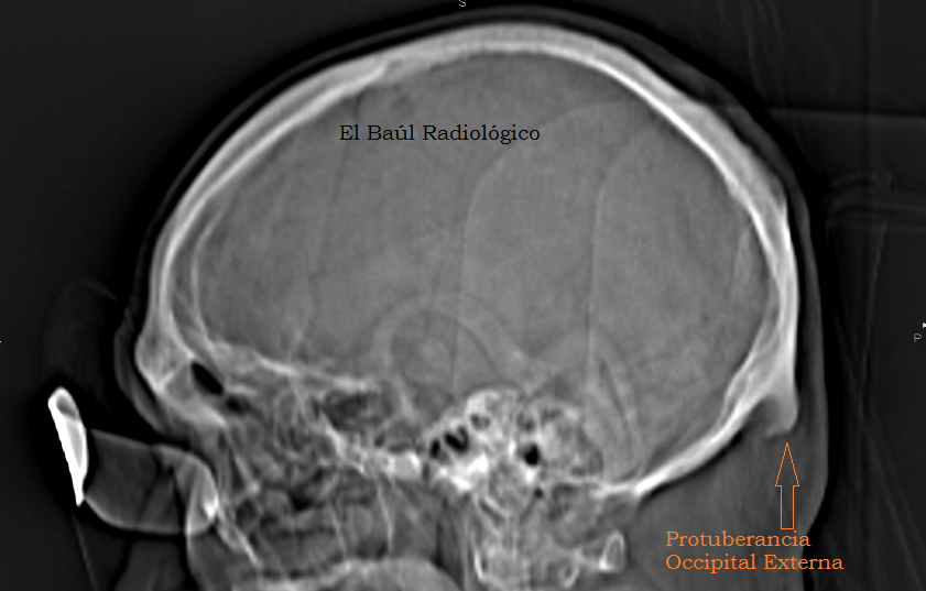
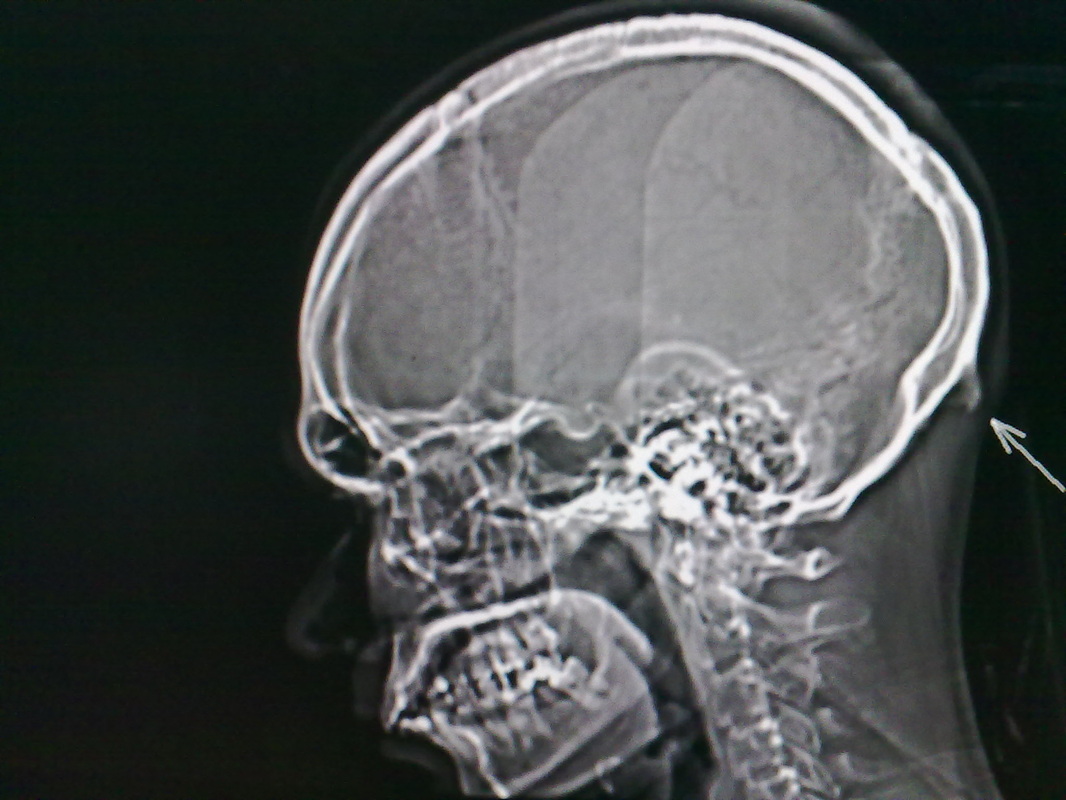
Case Discussion. External occipital protuberance is a midline bony prominence in the occipital bone that ligamentum nuchae and trapezius muscle attach to its tip. The tentorium cerebelli attaches to its internal surface. Exaggerated external occipital protuberance also is known as an occipital spur.

El Baúl Radiológico PROTUBERANCIA OCCIPITAL EXTERNA (Ossified Spur on the External Occipital
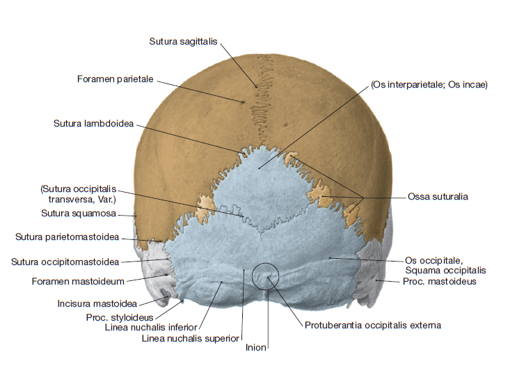
Protuberantia occipitalis externa Read more. Description. The external occipital protuberance is the palpable prominence found along the external aspect of the squamous part of occipital bone. It is located at the point along the midline where the occipital and nuchal planes meet. It consists of the inion, which is a craniometric point located.

Protuberantia occipitalis externa MedkoM
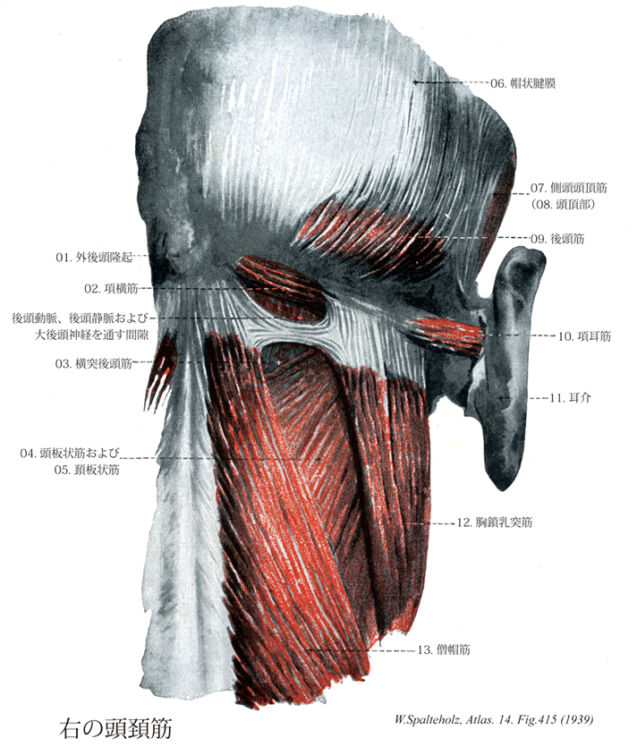
Die Protuberantia occipitalis externa dient als Ursprung des Ligamentum nuchae und des am weitesten kranial gelegenen Teils des Musculus trapezius. Sie markiert den Punkt Inion . Die Protuberantia occipitalis externa ist eine prominente Knochenvorwölbung in der Mitte der Außenfläche des Os occipitale.

El Baúl Radiológico PROTUBERANCIA OCCIPITAL EXTERNA (Ossified Spur on the External Occipital
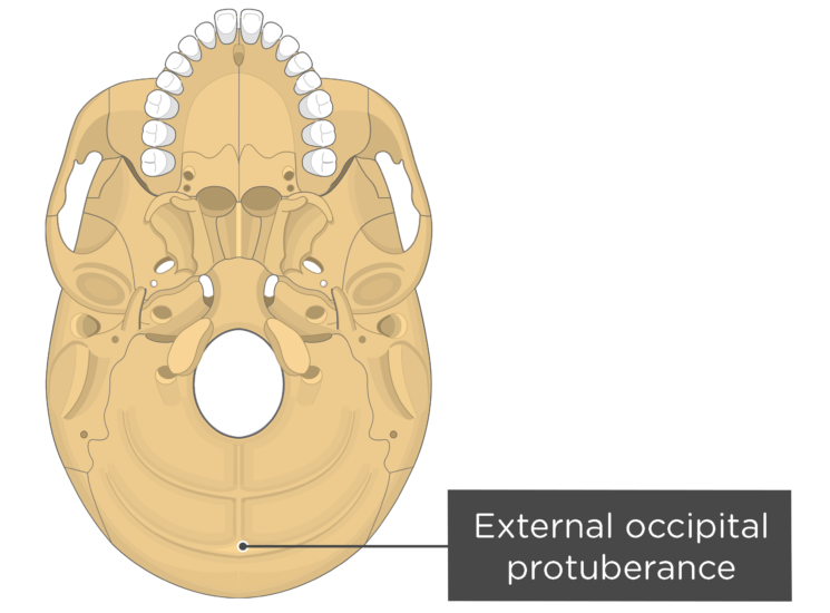
Protuberantia occipitalis externa. Definition. The under surface of the jugular process is rough, and gives attachment to the Rectus capitis lateralis muscle and the lateral atlantoöccipital ligament; from this surface an eminence, the paramastoid process,sometimes projects downward, and may be of sufficient length to reach, and articulate.
Occipital Bone Markings
Introduction: In controversial fashion, the presence of an enlarged external occipital protuberance has been recently linked to excessive use of handheld electronic devices.We sought to determine the prevalence of this protuberance in a diverse age group of adults from two separate time periods, before and approximately 10 years after the release of the iPhone, to further characterize this.

External Occipital Protuberance Earth's Lab
Near the line where it unites to the interparietal bone, the squamous part of the occipital bone shows the transversal prominence of the external occipital protuberance (protuberantia occipitalis externa). In species like Dogs or Horses, where the occipital reflexes on the dorsal surface of the head, this protuberance occupies the upmost point of the cranium and marks the dorsal elbow of the.
Protuberance Anatomy Anatomical Charts & Posters
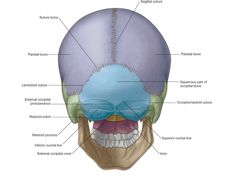
The occipital squama is the larger, more posterior portion of the external occipital bone. It is a thin, flat bone that forms the base of the skull and contributes to the formation of the posterior cranial fossa.The occipital squama is marked by several prominent features, including the external occipital protuberance, the external occipital crest, and the superior nuchal lines.
Terminologia Anatomica(TA)に基づく解剖学
External Occipital Protuberance (EOP) enlargement has been recently reported to increase in young adults, with a putative link with postural factors such as the use of smartphones. This study aims.
Protuberanta occipitala externa Despre viața din România
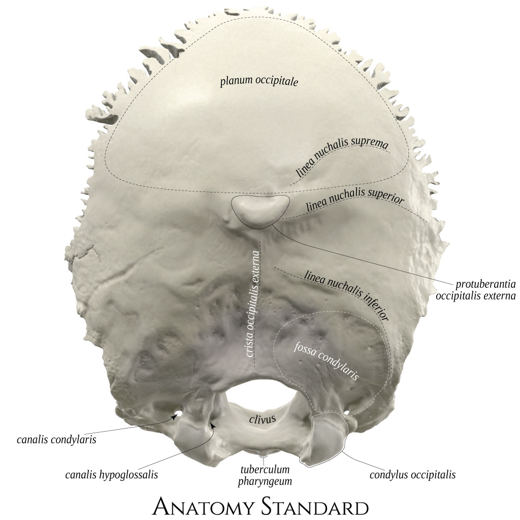
Protuberantia occipitalis externa. Crista occipitalis externa. Fossa condylaris. Condylus occipitalis. Pars basilaris ossis occipitalis. Tuberculum pharyngeum. Canalis nervi hypoglossi. Canalis condylaris. Clivus. Uploaded by: rva Netherlands, Leiden - Leiden University Medical Center, Leiden University.
Anatomy Standard Drawing Occipital bone exterior (posterior) view Latin labels AnatomyTOOL
Protuberantia occipitalis externa Clavicula Acromion Spina scapulae Angulus inferior Plica axillaris posterior Musculus erector spinae Os coxae Os sacrum Os coccygis Tuber ischiadicum Palpable bony structures 11071-01_CH01.qxd 3/28/08 10:05 AM Page 4. PLATE 1-03 PAGE 7 Chapter 1 Lippincott Williams & Wilkins Atlas of Anatomy

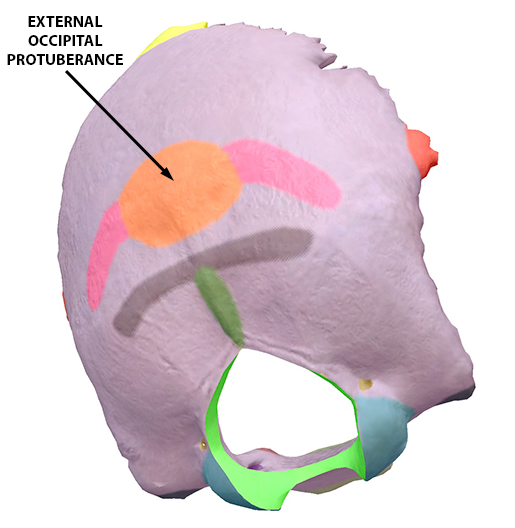
External occipital protuberance a site for attachment of muscles and ligaments Body bones
External occipital protuberance (EOP) is a midline bony prominence in the occipital bone that ligamentum nuchae and trapezius muscle attach to its tip that named Inion. The tentorium cerebelli attaches to its internal surface. Entheses are the sites of ligament, tendon or joint capsule attachment to the bone.
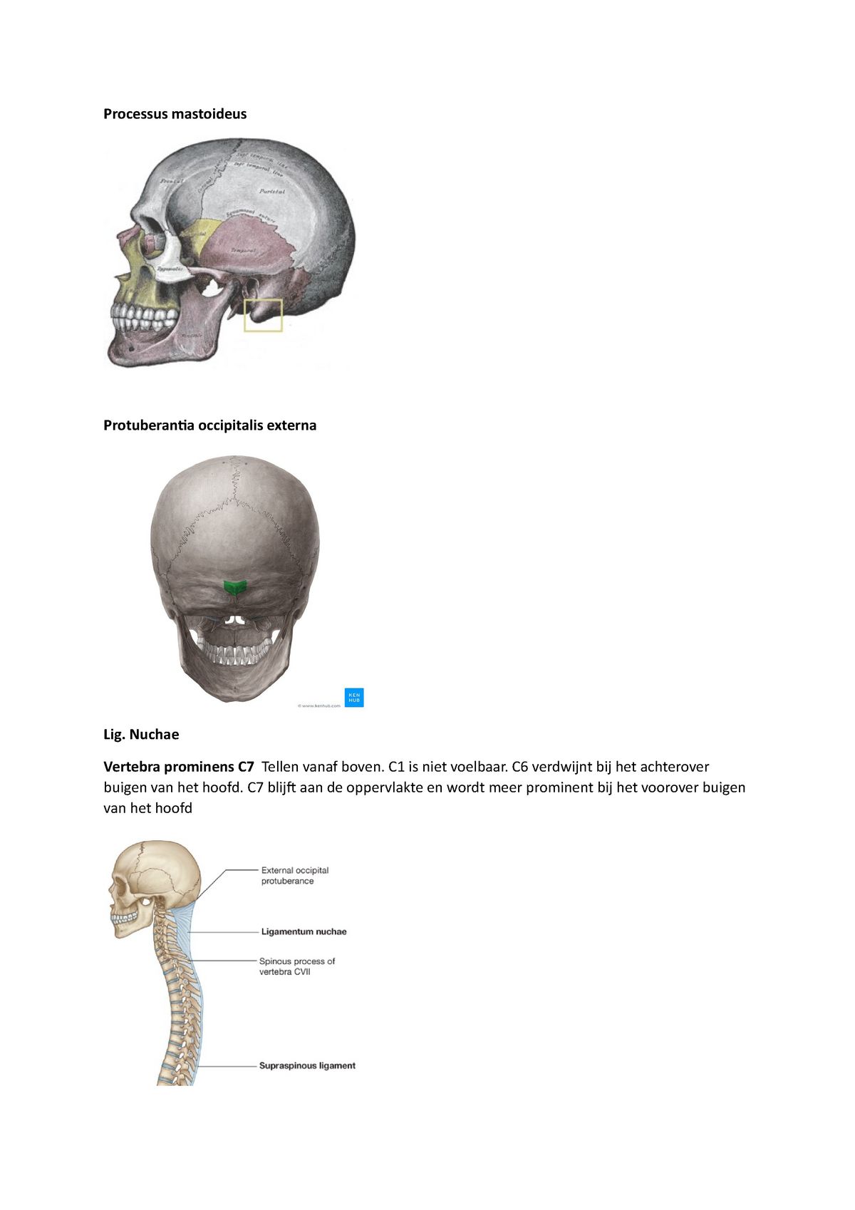
Anatomie lijst met figuren Processus mastoideus Protuberantia occipitalis externa Lig. Nuchae
external occipital protuberance: [TA] a prominence about the center of the outer surface of the squamous portion of the occipital bone, giving attachment to the ligamentum nuchae. Synonym(s): protuberantia occipitalis externa [TA]
El Baúl Radiológico PROTUBERANCIA OCCIPITAL EXTERNA (Ossified Spur on the External Occipital
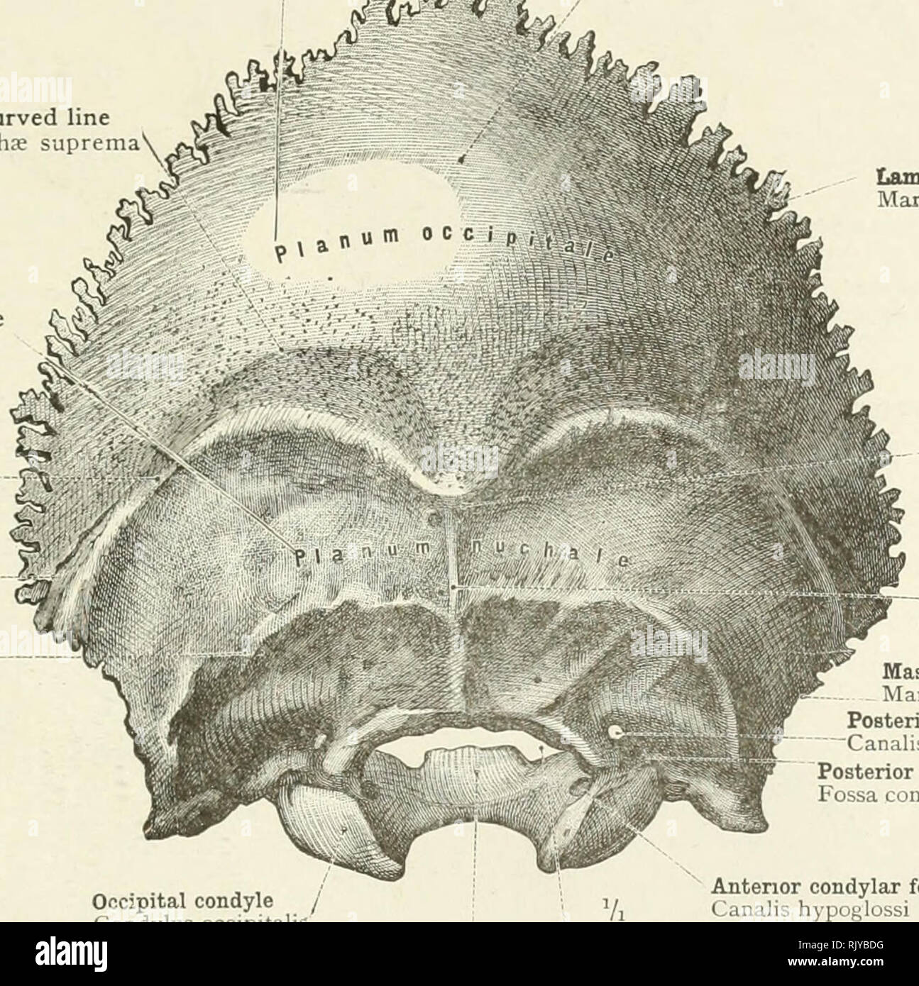
Dorsal aspect of occipital bone. The prominent feature of the squama's external surface is the protuberance, an easily palpable point known in the craniometry as inion. The list of terms: Planum occipitale - Occipital plane Protuberantia occipitalis externa - External occipital protuberance Linea nuchalis suprema - Highest nuchal line Linea nuchalis superior - Superior nuchal line
Prominent External Occipital Protuberance Radiology
Protuberantia occipitalis externa - External occipital protuberance Linea nuchalis superior - Superior nuchal line Linea nuchalis inferior - Inferior nuchal line Crista occipitalis externa - External occipital crest Foramen magnum. That illustration shows canals of the base of the skull, which usually is more challenging to localize.

Vom Skelett zum Geschlecht Miss Jones
Epidemiology. It is common in males and hence is often used in forensic investigations for gender determination 1.. Clinical presentation. It is an anatomical variant which is usually noticed incidentally radiographically, although it can become symptomatic with affected patients describing a tender bony swelling at the back of the neck causing pain especially while lying down 1.
