
Anatomy Of Dog Skeleton With Labeled Inner Bone Scheme Vector
A major part of a dog's anatomy is their musculature. This is a system formed by muscles, tendons and ligaments. A dog can have between 200 and over 400 muscles.Again, the amount of muscles an individual dog has depends on the breed and the individual.

canine muscular anatomy Dog Muscles Diagram
Here, I will provide a dog skeleton labeled diagram and the different parts of a dog diagram. In the dog skeleton labeled diagram, I tried to show you all the bones from the body. This might help you understand the different regions of the body so quickly. I would like to show different external features of a dog again here in a labeled picture.
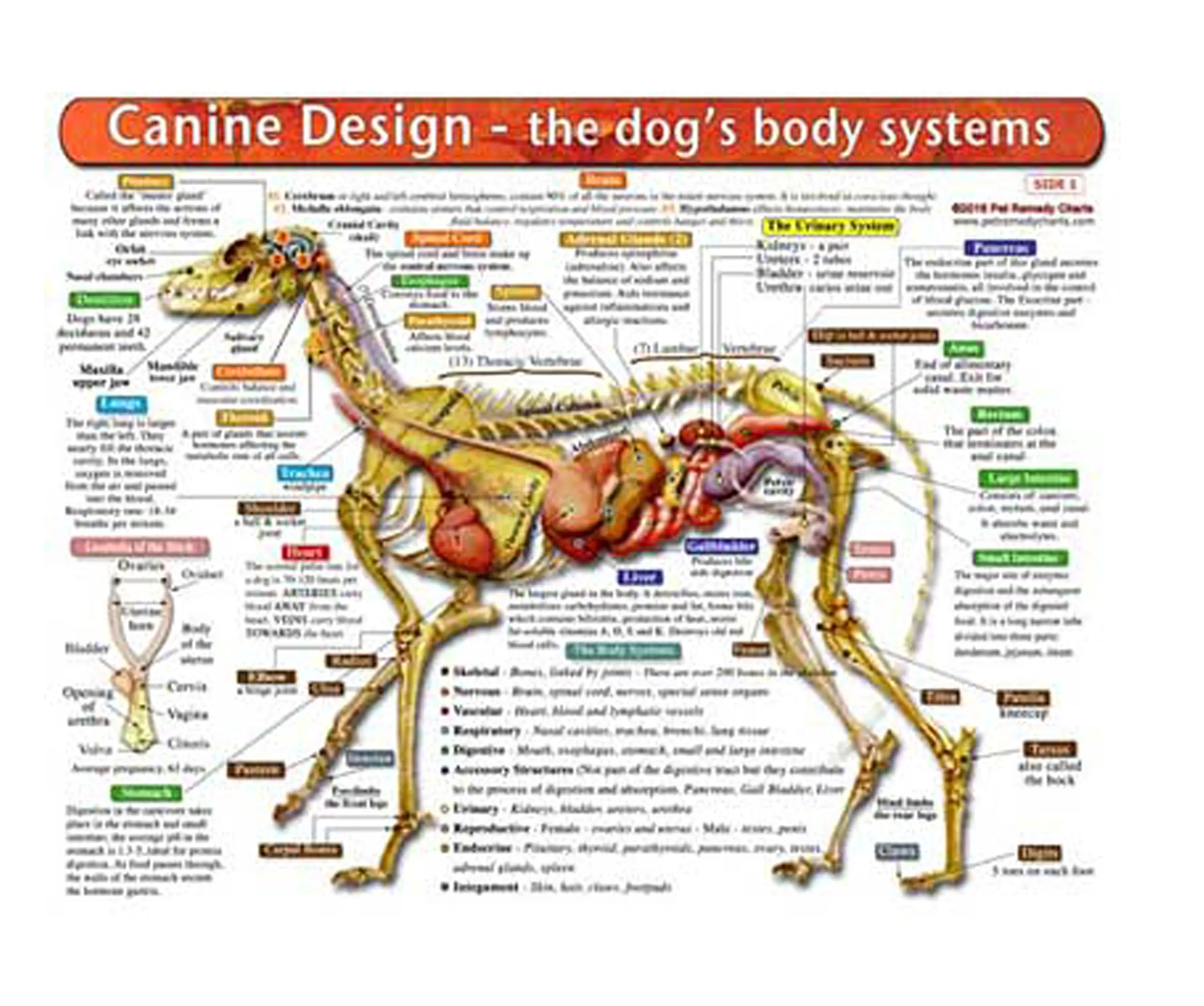
Buy The Dogs Body Systems A DoubleSided, UV Protected, Laminated Dog
The vertebrae of the dog skeleton anatomy also possess some exceptional osteological features that a ruminant or horse. Now, you should try to identify all the bones from the dog skeleton from the actual samples of your anatomy laboratory. You may use the dog bones labeled diagram from the anatomy learner.

an image of a dog's muscles labeled in the body and labelled with names
Skeleton of the dog. Axial Skeleton. = the Head and spinal column. Head - made up of around 50 bones. Skull - occipital crest along top, occiput - rear point of skull zygomatic arch - under and around the base of the eye. Muzzle - nasal crest (largely cartilaginous) top of nose. - maxilla - top jaw. mandible - bottom jaw.
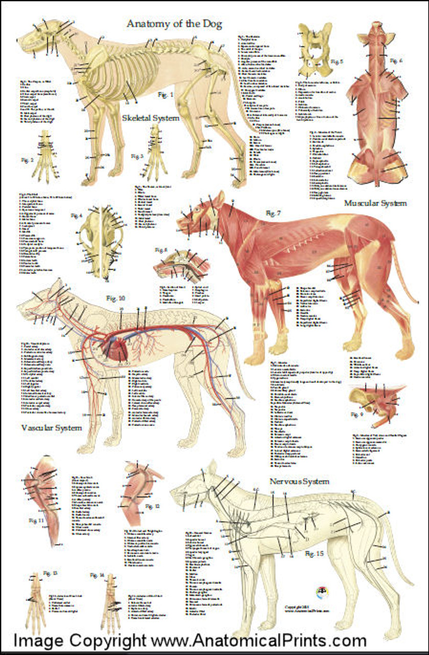
M. Douglas Wray Dog Anatomy
Anatomic Planes. The main planes of motion for dogs are as follows (see Figure 5-1): • The sagittal plane divides the dog into right and left portions. If this plane were in the midline of the body, this is the median plane or median sagittal plane. • The dorsal plane divides the dog into ventral and dorsal portions.
Dog Anatomy Poster 24 x 36 Clinical Charts and Supplies
A dog's skeleton is made up of many different bones, which provide structure and support for their body. Dogs have over 300 bones in their body, which is more than humans who have around 206 bones. Their skeleton includes their skull, spine, ribcage and limbs. Dogs have four legs that are designed to help them move quickly and efficiently.

Anatomy of dog with inside organ structure examination vector
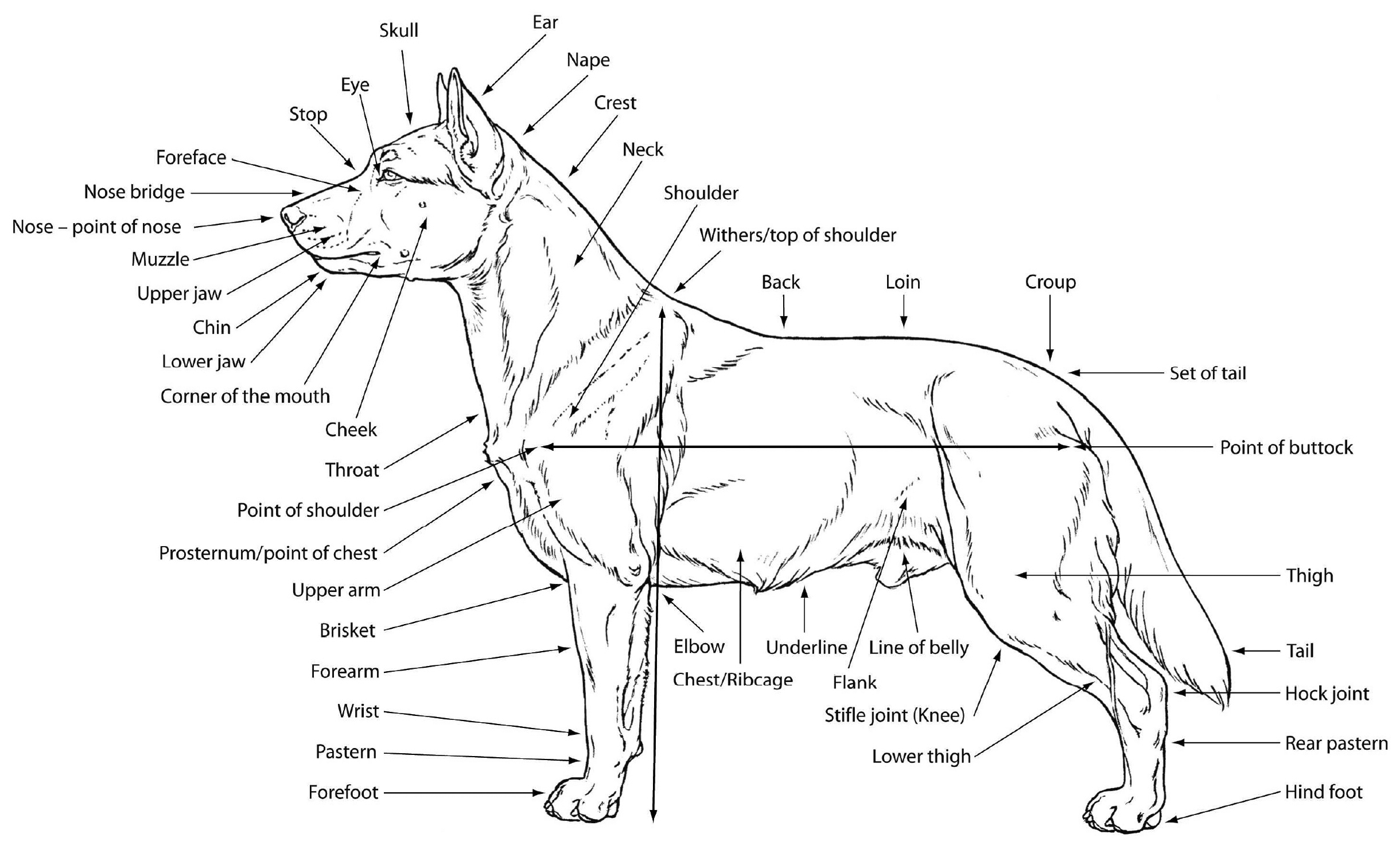
Head's up on dog parts. Starting from the head, a dog is made up of the. Nose: Dog noses are often cold and wet, and of course, they usually get stuck where they're not wanted. The muzzle (foreface) comprised of the upper and lower jaws. The stop is an indentation (sometimes nonexistent) between the muzzle and the braincase or forehead.
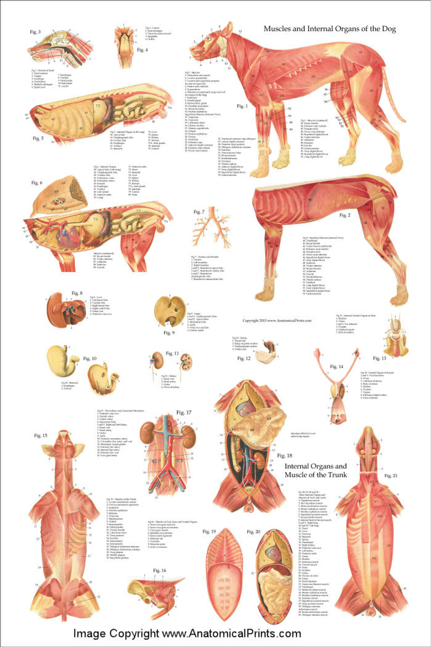
Dog Muscles And Internal Anatomy Chart Clinical Charts and Supplies
A female dog's reproductive system has similar organs as a human's. The female dog anatomy external organ is the vulva, which opens to the vagina. A pregnant female dog's anatomy includes two ovaries, which produce eggs, the cervix, fallopian tubes, and the uterus. The uterus becomes the womb for her puppies during their gestation period.

PetMassage™ Chart 5 Superficial Muscles of the Dog · PetMassage
This module of vet-Anatomy is a basic atlas of normal imaging anatomy of the dog on radiographs. 51 sampled x-ray images of healthy dogs performed by Susanne AEB Borofka (PhD - dipl. ECVDI, Utrecht, Netherland) were categorized topographically into seven chapters (head, vertebral column, thoracic limb, pelvic limb, larynx/pharynx, thorax and abdomen/pelvis).

Dog Anatomy Dog Skelton
Anatomy atlas of the canine general anatomy: fully labeled illustrations and diagrams of the dog (skeleton, bones, muscles, joints, viscera, respiratory system, cardiovascular system). Positional and directional terms, general terminology and anatomical orientation are also illustrated.

Глубокие мышцы, внутренние органы собаки Dog Muscles & Internal
Dog anatomy comprises the anatomical studies of the visible parts of the body of a domestic dog.Details of structures vary tremendously from breed to breed, more than in any other animal species, wild or domesticated, as dogs are highly variable in height and weight. The smallest known adult dog was a Yorkshire Terrier that stood only 6.3 cm (2.5 in) at the shoulder, 9.5 cm (3.7 in) in length.

dogexternalanatomy ESL Buzz
These include the head, ears, eyes, nose, mouth, neck, tail, legs, and paws. The head of a dog is one of its most distinguishing features. It includes the skull, jaw, and teeth. The ears can be upright or floppy, and they come in a variety of shapes and sizes. The eyes are usually round and can be brown, blue, or green.

PetMassage Chart 3 Skeleton of the Dog · PetMassage™ Training and
It provides information about a dog's skeletal, reproductive, internal, and external anatomy, along with accompanying labeled diagrams. After mating, dogs experience something called a copulatory tie, wherein they remain in the coital position. The male dog dismounts the female at this time. The dogs can remain in this position from a few.

Dog neck anatomy
The Anatomage Table Vet brings realism into veterinary education by providing an absolutely explicit 3D visualization of animal anatomy. The platform features 3D animal bodies - which include the world's most detailed canine cadaver - that assist anatomy inspection. Introduce a high-quality approach to animal learning using digital.

Parts of a Dog Useful Dog Anatomy with Pictures • 7ESL
Whereas giant breeds can take between 18 months and 2 years for their growth plates to fuse. Speaking of skeletons, a dog has 320 bones in their body (depending on the length of their tail) and around 700 muscles. Muscles attach to bones via tendons. Depending on the breed of dog, they will have different types of muscle fibers.

dog anatomy Dog Care Training Grooming
This veterinary anatomy module contains 608 illustrations on the canine myology. Here are presented scientific illustrations of the canine muscles and skeleton from different anatomical standard views (lateral, medial, cranial, caudal, dorsal, ventral / palmar.). Some fascias, tendons, ligaments, joints were labeled.
