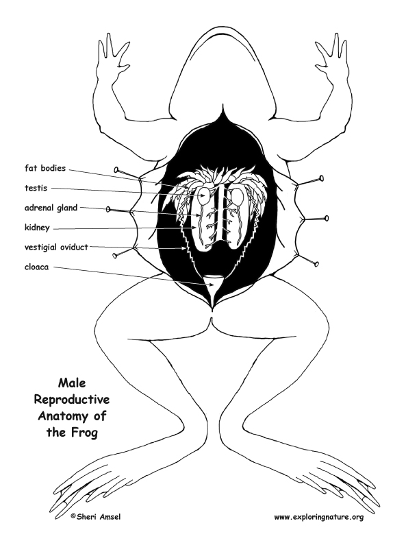
Frog Reproductive Anatomy Diagram and Labeling
How To Draw A Frog | Labeled Diagram Of Frog - YouTube © 2023 Google LLC #frog #frogdiagram #howtodrawStudents need to learn about the basic parts of a frog. So in this video, I try to.
All about frogs and toads Wildlife
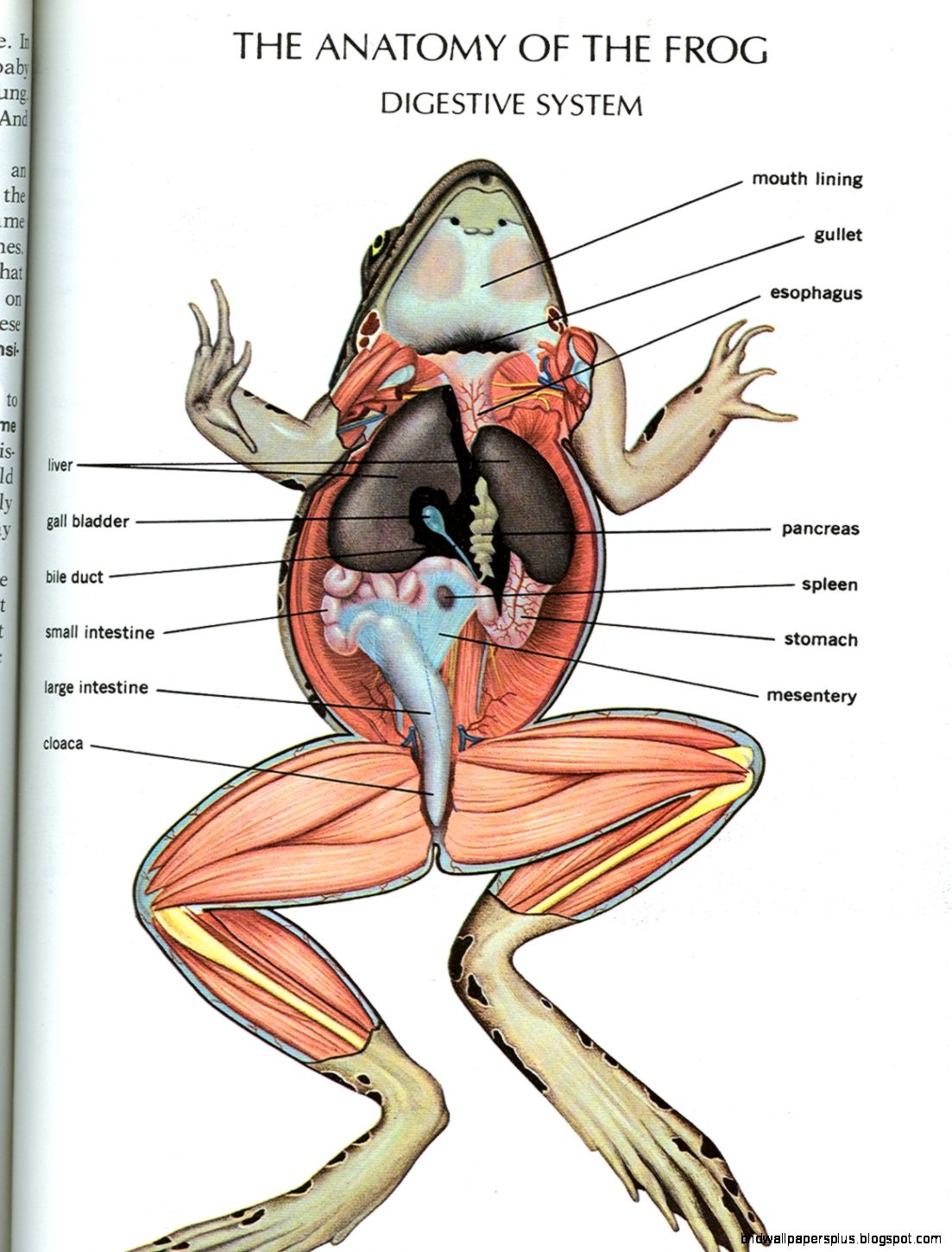
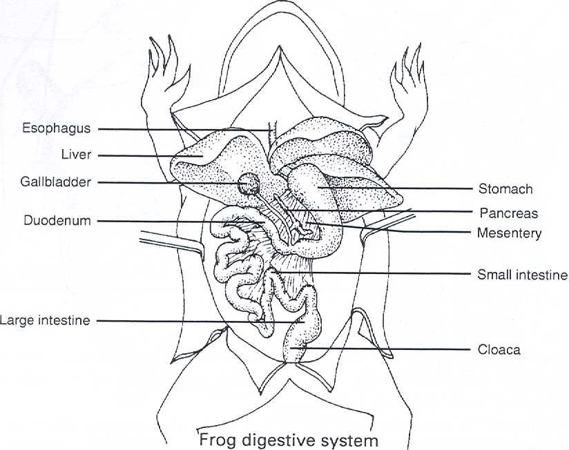
This BiologyWise post provides a labeled frog digestive system diagram to help you understand the digestive process in frogs. After catching their prey, frogs close their eyes and retract them through the holes in their skull. This helps them push food down the throat.
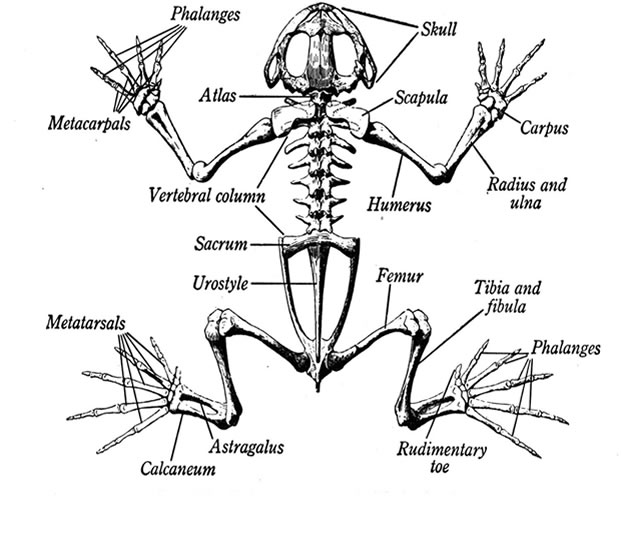
Skeletal Anatomy of a Frog Bones Within A Frog
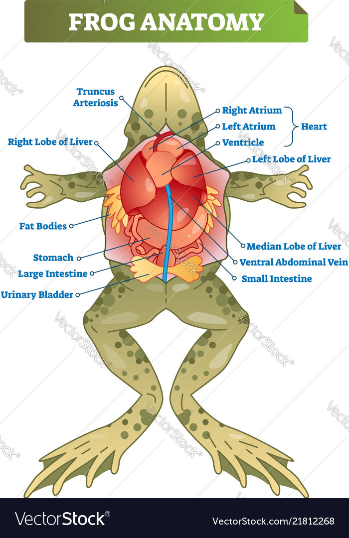
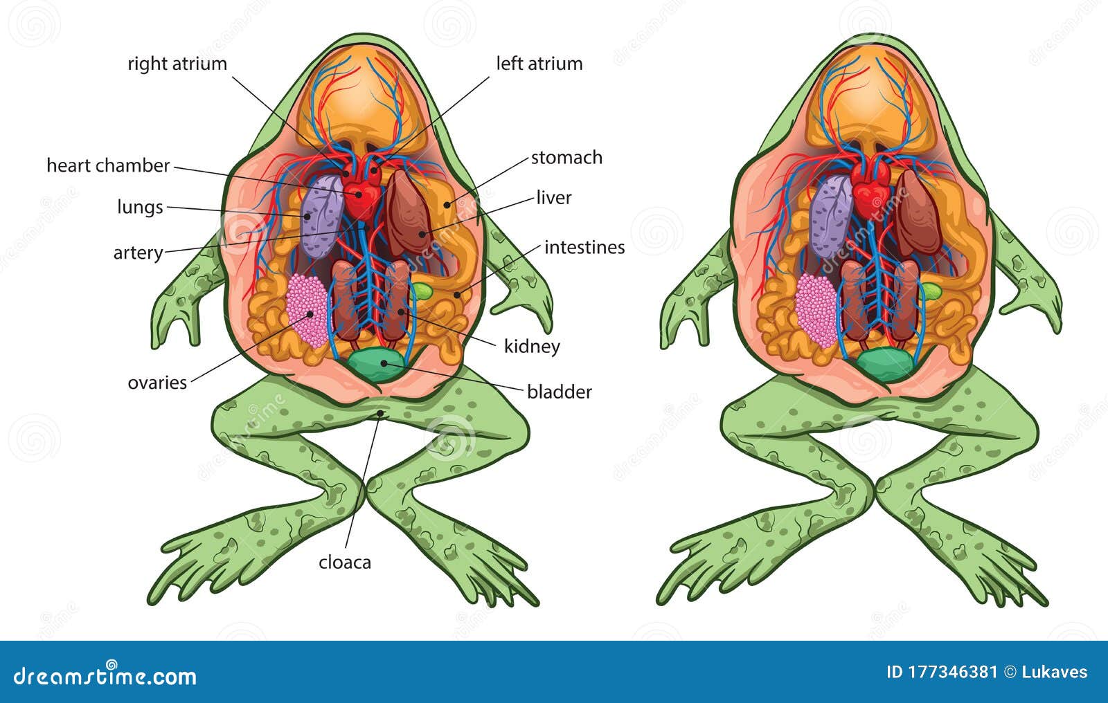
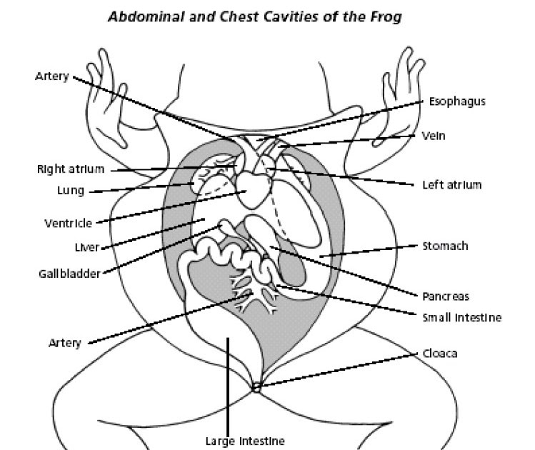
Frog Anatomy Label This worksheet is a supplement to the frog dissection activity where students examine a preserved specimen. The main structures of the abdominal cavity are shown on this image and students practice identifying them using the included word bank.
Frog Anatomy HD Wallpapers Plus
Procedure: Put on safety goggles, gloves, and a lab apron. Place a frog on a dissection tray. To determine the frog's se x, look at the hand digits, or fingers, on its forelegs. A male frog usually has thick pads on its "thumbs," which is one external difference between the sexes, as shown in the diagram below.

Parts of a Frog Nomenclature Cards
Refer to the interactive diagram above to learn where each part is located. Maxilla - Forms the upper jawbone Atlast - The top part of a backbone Suprascapula - Shoulder blade Vertebrae - Individual bones that form the spine Sacral Vertebra - A bone below the last vertebra, positioned between the hips
Frog anatomy labeled scheme Royalty Free Vector Image
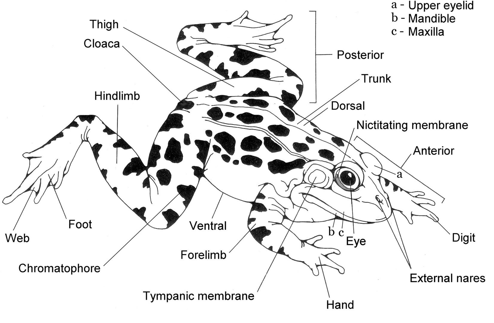
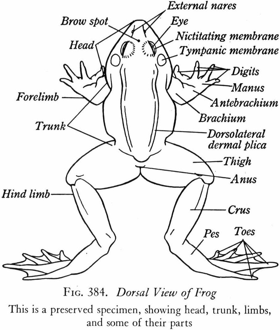
Why not start by labelling this diagram. This exercise is for students in 1st, 2nd, 3rd, 4th, 5th, 6th and 7th grades. A well labelled diagram of a frog and toad Here is a description of the function of each part of the frog: Head - contains the brain, which controls the body's functions and sensory organs
Frog Dissection External Anatomy
Tadpoles or Polliwogs are the aquatic larval stage of frogs that evolved from eggs after 3 to 25 days. They measure about 40-45mm and live in water. Tadpoles evolve for 14 to 16 weeks depending on the species and the climate in which they live. Once frog eggs have hatched, they will turn into tadpoles.
Frog anatomy stock vector. Illustration of population 177346381
Today I will show you " How to draw and label diagram of frog easily step by step | How to draw frog ".

The Frog's Anatomy Illustration Poster Graphic poster
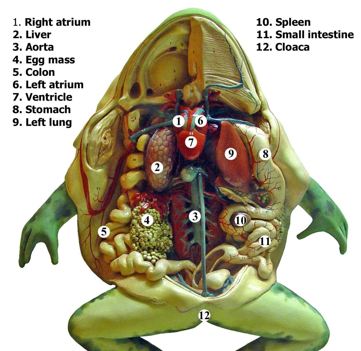
Heart. The frog's heart is the small triangular organ at the top. Unlike a mammal heart, it only has three chambers — two atria at the top and one ventricle below. Carefully cut away the pericardium, the thin membrane surrounding the heart. Notice the arteries connected to the top of the heart, giving it a 'Y' shape.
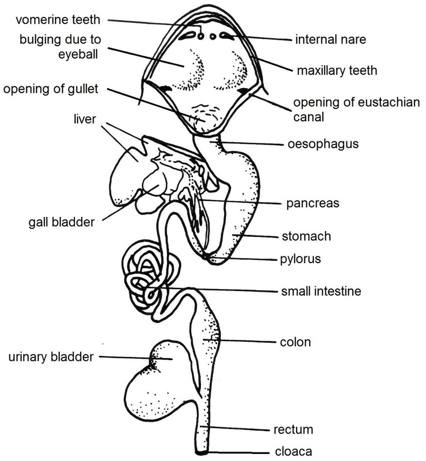
Digestive system of frog Anatomy and Physiology of digestion Online
How To Draw A Frog Very Simple & Easy | Labelled Diagram Of Frog | Biology Diagram - YouTube © 2023 Google LLC In this you are going to learn how to draw labelled diagram of Frog.
Frog Dissection MRS. MERRITT'S BIOLOGY CLASS
5. Color and Label the Organs of the Frog 6. Internal Anatomy of the Frog with Liver Removed Diagram (Color) 7. Internal Anatomy of the Frog with Liver Removed Labeling (Color) 8. Internal Anatomy of the Frog with Liver Removed Diagram (BW) 9. Internal Anatomy of the Frog with Liver Removed Labeling (BW) 10. Comparing the Anatomy of the Frog.
External Anatomy Of A Frog Anatomical Charts & Posters
The style of citing shown here is from the MLA Style Citations (Modern Language Association). When citing a WEBSITE the general format is as follows. Author Last Name, First Name (s). "Title: Subtitle of Part of Web Page, if appropriate." Title: Subtitle: Section of Page if appropriate. Sponsoring/Publishing Agency, If Given.
Frog Pre Lab/Lab Core 71 Science
Run you finger over both sets of teeth and note the differences between them. 6. On the roof of the mouth, you will find the two tiny openings of the nostrils, if you put your probe into those openings, you will find they exit on the outside of the frog. 7. Label each of the structures underlined above. 8.

a frog with its mouth open and tongue out
Frog labeled diagram drawing / How to draw and label Frog diagram Biology / Science Projects CBSEIn this video, I will learn How to draw and label Frog diagr.

Parts of a frog Grammar Tips
Animal Diagrams: Frog (labeled and unlabeled) Overview. Diagram of a frog. Media PDF. Download Resource Tags. Amphibians Animal Diagrams Frog & Toads. Similar Resources PREMIUM. Paper Bag Puppet: Animals - Tree Frog / Paper Bag Puppets. Media Type PDF. PREMIUM. Animal Diagrams: Chrysalis (unlabeled parts)
LLA BIOLOGY Simple Frog Diagram
3. Examine the inside of the mouth. Use your scalpel to cut the membrane that connects the hinges of the frog's mouth and open the mouth widely to examine the inside. You should be able to see and label the esophagus, which connects to the stomach, and the glottis, which connects to the lungs.
