
Os temporale Ars Neurochirurgica
The petrous part (also called the pyramid) is the part of the temporal bone which houses the inner ear. It is located in the base of the skull between the sphenoid and occipital bones.
Anatomy Standard Drawing Temporal bone medial view Latin labels AnatomyTOOL
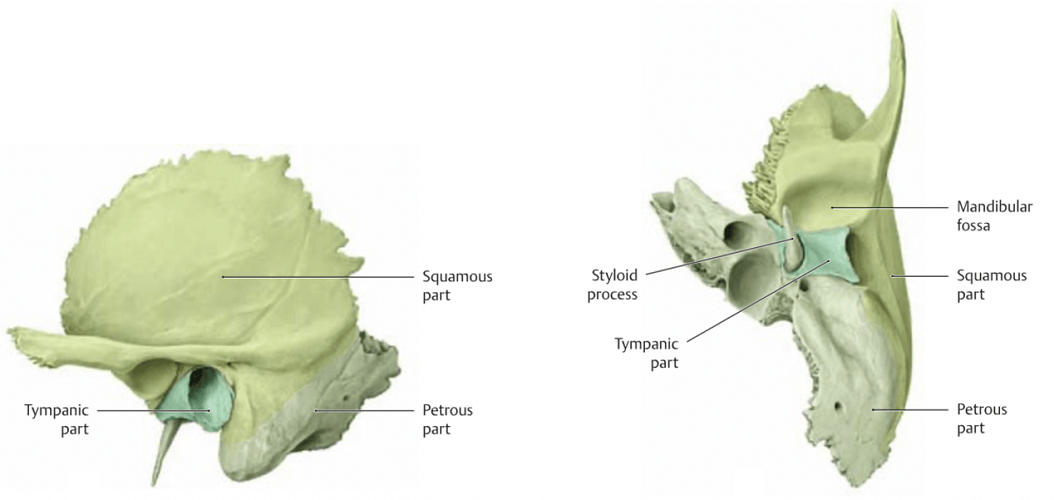
In this pdf-note, we will learn about the structure of the temporal bone (os temporale). It contains the organs of hearing and balance, and many blood vessels and nerves pass through it. Temporal bone (os temporale) There are several parts of the temporal bone: Petrous part (pars petrosa) Petrous part (pars petrosa) Tympanic part (pars tympanica)

Os temporale Ars Neurochirurgica
. Internal acousttic meatus and the osseous labyrinth within the temporal bone The red structure you see in the image above is the osseous labyrinth - the channel system surrounded by a thick cortical bone layer within the petrous part of the temporal bone.
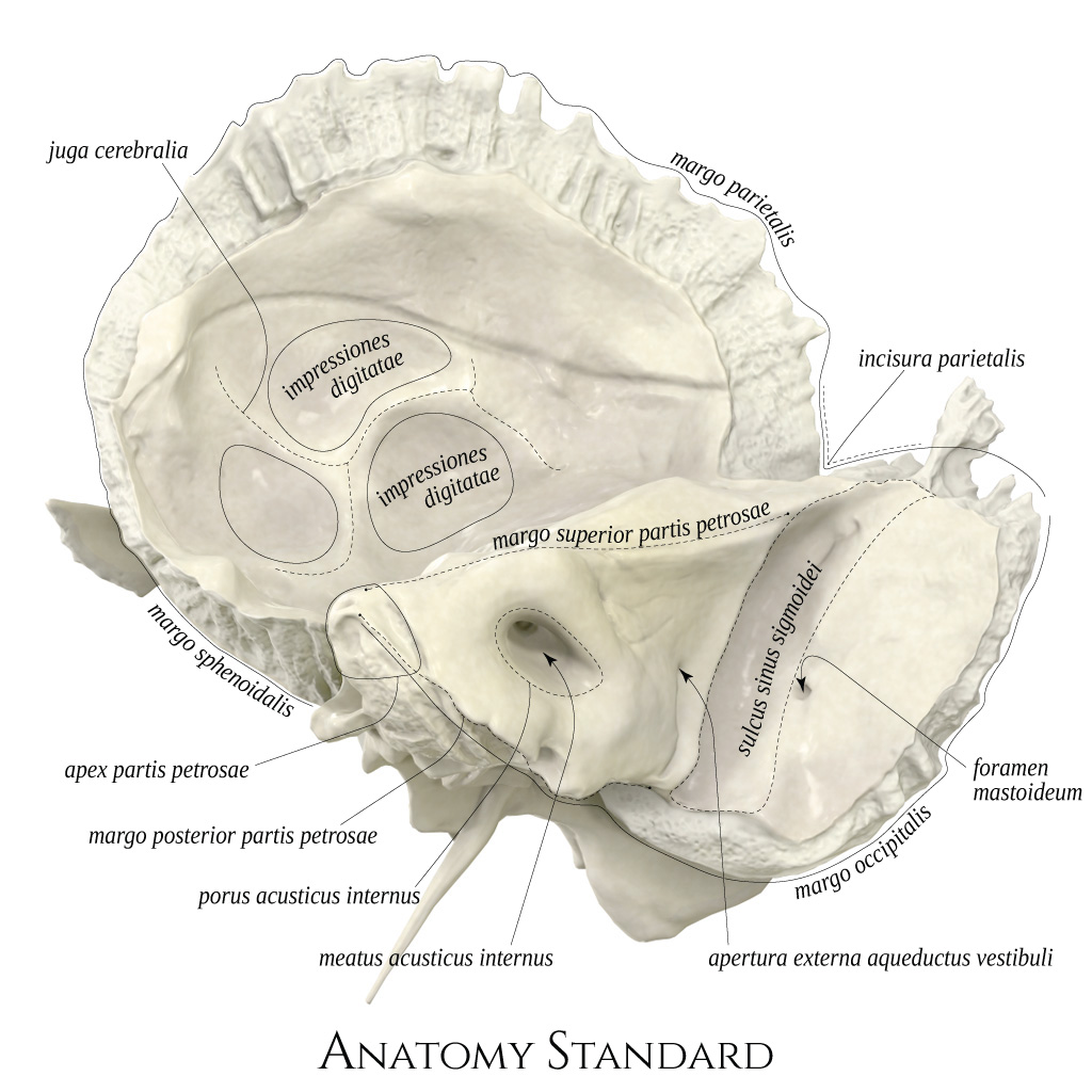
Anatomy Standard Drawing Temporal bone superior view Latin labels AnatomyTOOL
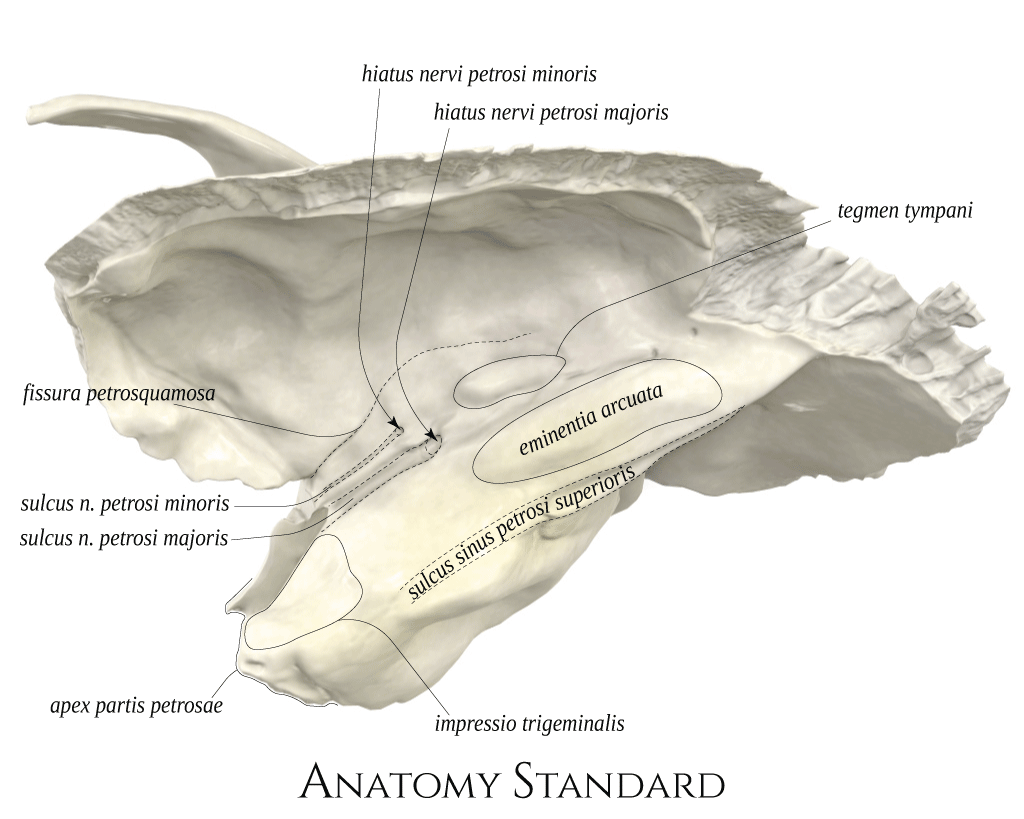
Pars petrosa ossis temporalis Latin synonym: Pyramis; Pars petromastoidea Synonym: Petrous bone; Pyramis Acronym: PTB Related terms: Petrous part; Petrous part (Temporal bone); Temporal bone: Petrous part Definition The petrous part or pyramid is pyramidal and is wedged in at the base of the skull between the sphenoid and occipital.

Os temporale Axon
The petrous part of the temporal bone, also known as the petrous temporal bone ( PTB ), forms the part of skull base between the sphenoid and occipital bones. Gross anatomy The petrous temporal bone has a pyramidal shape with an apex and a base as well as three surfaces and angles: apex ( petrous apex)

Os temporale Axon
3 The temporal bone (Os temporale) consists of the petrous portion (Pars petrosa), the. tympanic part (Pars tympanica), the squama temporalis part (Pars squamosa) and the.
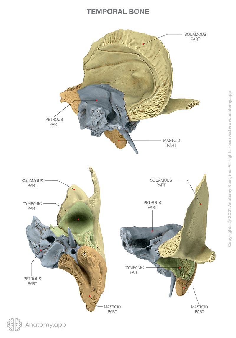
Temporal bone Encyclopedia Anatomy.app Learn anatomy 3D models, articles, and quizzes
This protocol describes how to obtain bone powder from the pars petrosa of disarticulated ossis temporalis, specifically from the dense parts around the cochlea, in a minimally-invasive way by.

Minimallyinvasive sampling of pars petrosa (os temporale) for ancient DNA extraction
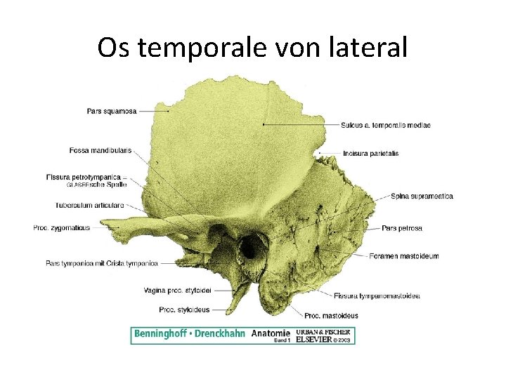
Outer surface. (Os Temporale) The temporal bones are situated at the sides and base of the skull. Each consists of five parts, viz., the squama, the petrous, mastoid, and tympanic parts, and the styloid process. The Squama (squama temporalis). —The squama forms the anterior and upper part of the bone, and is scale-like, thin, and translucent.

pars petrosa ossis temporalis Tıpacı
Anatomical structure of pars petrosa of os temporale sinister. (a) MAE, Meatus acusticus externus. (b) Paramedian section of the pars petrosa between osseous external ear canal and middle ear structure; ME, middle ear with the tympanic cavity proper (---); Nf, nervus facialis; OEEC, osseous external ear canal.
Temporal Bone The Definitive Guide Biology Dictionary
This protocol describes how to obtain bone powder from the pars petrosa of disarticulated ossis temporalis, specifically from the dense parts around the cochlea, in a minimally-.

Petrous Part of Temporal Bone ParisrilloFrye
Annotated and non-annotated images are provided in this temporal bone teaching CT.
Anatomy Standard Drawing Temporal bone lateral view Latin labels AnatomyTOOL
Category:Petrous part of the temporal bone From Wikimedia Commons, the free media repository Media in category "Petrous part of the temporal bone" The following 20 files are in this category, out of 20 total. Alpine ibex petrol bone.jpg 3,331 × 3,006; 3.96 MB Base of skull 17.jpg 960 × 720; 108 KB Braus 1921 343.png 1,536 × 1,092; 4.81 MB
Os temporale (Schläfenbein) Anatomie, Lage und Suturen Kenhub
Mittlerer Teil (pars sellaris): •Corpus ossis sphenoidalis Lat. Teilen (p. temporalis): •Ala major ossis sphenoidalis •Os temporale: p. squamosa, p. Petrosa facies ant. Borders : anterior: •Ala minor, •Sulcus prechiasmatis posterior: •Dorsum sellae •Margo sup. partis petrosae
Os temporale (Schläfenbein) Anatomie, Lage und Suturen Kenhub
Pars squamosa ossis temporalis 1/4 Synonyms: none The squamous part is the anterior superior portion of the temporal bone that forms the lateral part of the middle cranial fossa. It has the appearance of a large flattened plate. Its external surface is smooth and slightly convex.
33 Os temporale uere und innere Schdelbasis Dvid
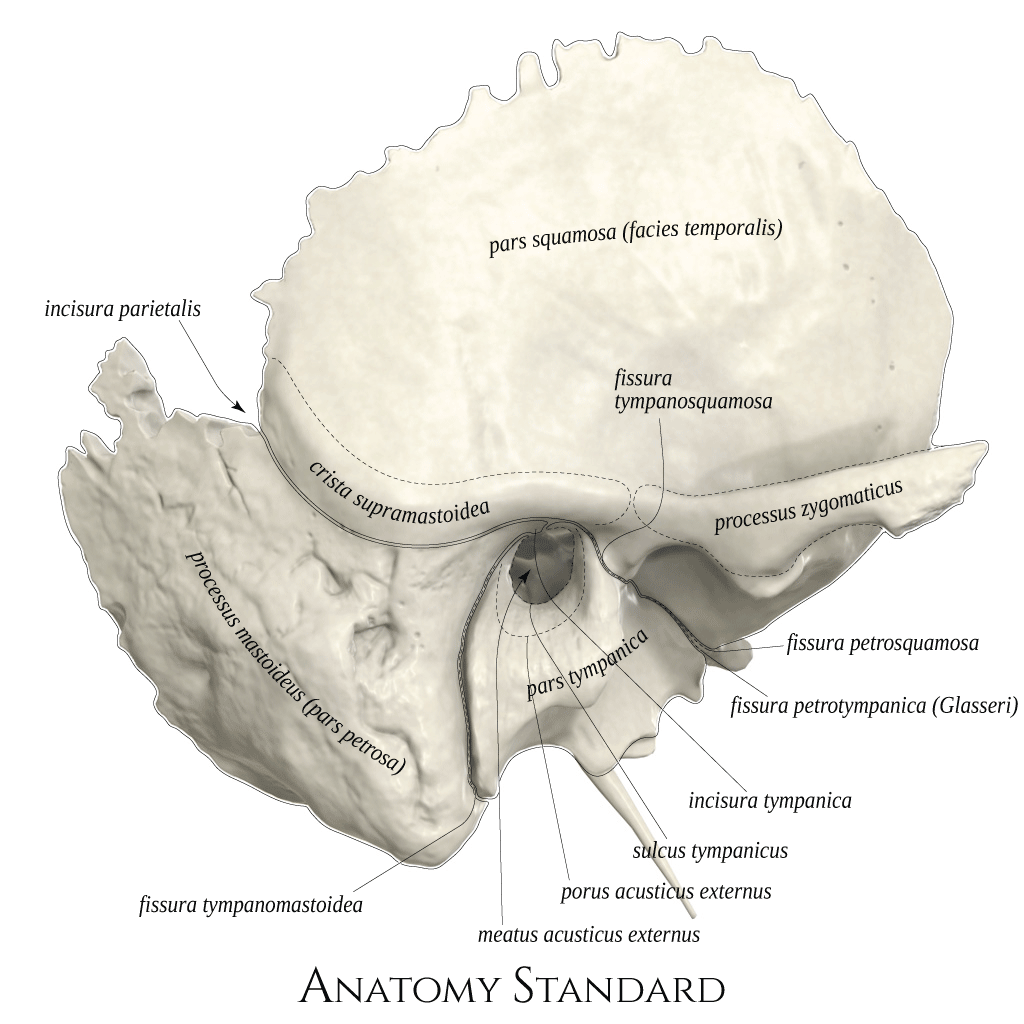
Additional formats:None available. Description: Temporal bone: lateral view. The temporal bone (os temporale) is shown from inferior. Latin labels. Image retrieved from Anatomy Standard. Anatomical structures in item: Os temporale. Pars squamosa ossis temporalis.

Os temporale Axon
DEC 11, 2020 Minimally-invasive sampling of pars petrosa (os temporale) for ancient DNA extraction V.2 In 2 collections Eleftheria Orfanou,Marie Himmel,Franziska Aron,
