
Pin on Veterinária
Aims: In this study, a non-pathological vascularization model of feline abdomen was conducted on three adult cats was using anatomical and diagnostic imaging techniques. Methods: A live pet cat and two cat cadavers were used in this study.

Cat Anatomy (Thoracic and Abdominal Organs)
Edit article Citation, DOI, disclosures and article data This article lists a series of labeled imaging anatomy cases by body region and modality. Brain CT head: non-contrast axial CT head: non-contrast coronal CT head: non-contrast sagittal CT head: non-contrast axial with clinical questions CT head: angiogram axial CT head: angiogram coronal

Cat musculature Atlas of Comparative Vertebrate Anatomy
June 16, 2021 | Issue: July/August 2021 Krysta Janas DVM Karen Tobias DVM, MS, DACVS Yakov Oskanov/shutterstock.com Exploratory laparotomy or celiotomy is commonly performed for diagnosis, treatment, or prognostication of traumatic, inflammatory, infectious, neoplastic, and congenital abdominal conditions.
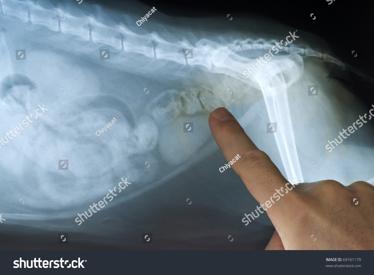
Cat Abdominal XRay Stock Photo 69161170 Shutterstock
25/04/2023 28/05/2022 by Sonnet Poddar The cat digestive system includes a mouth cavity, pharynx, alimentary canal, and different accessory organs. There are two major divisions in the mouth cavity of a cat - vestibule and mouth cavity proper. The alimentary canal of a cat starts with the esophagus and ends at the large intestine.
Torso Muscle Anatomy Diagram Biol 160 Human Anatomy And Physiology
We created an anatomical atlas of abdominal and pelvic CT which is an interactive tool for studying the conventional anatomy of the normal structures based on a multidetector computed tomography. Anatomical structures of the abdomen and pelvis are visible as interactive labeled images. Cross sectional anatomy: MDCT of the abdomen and pelvis

Muscles of the Abdomen and Ribs Laminated Anatomy Chart Anatomie
Atlas of CT Anatomy of the Abdomen This photo gallery presents the anatomy of the abdomen by means of CT (axial, coronal, and sagittal reconstructions). Click a link to get Axial view - Coronal view - Sagittal view < > Abdominal Computed Tomography

anatomy of the abdominals
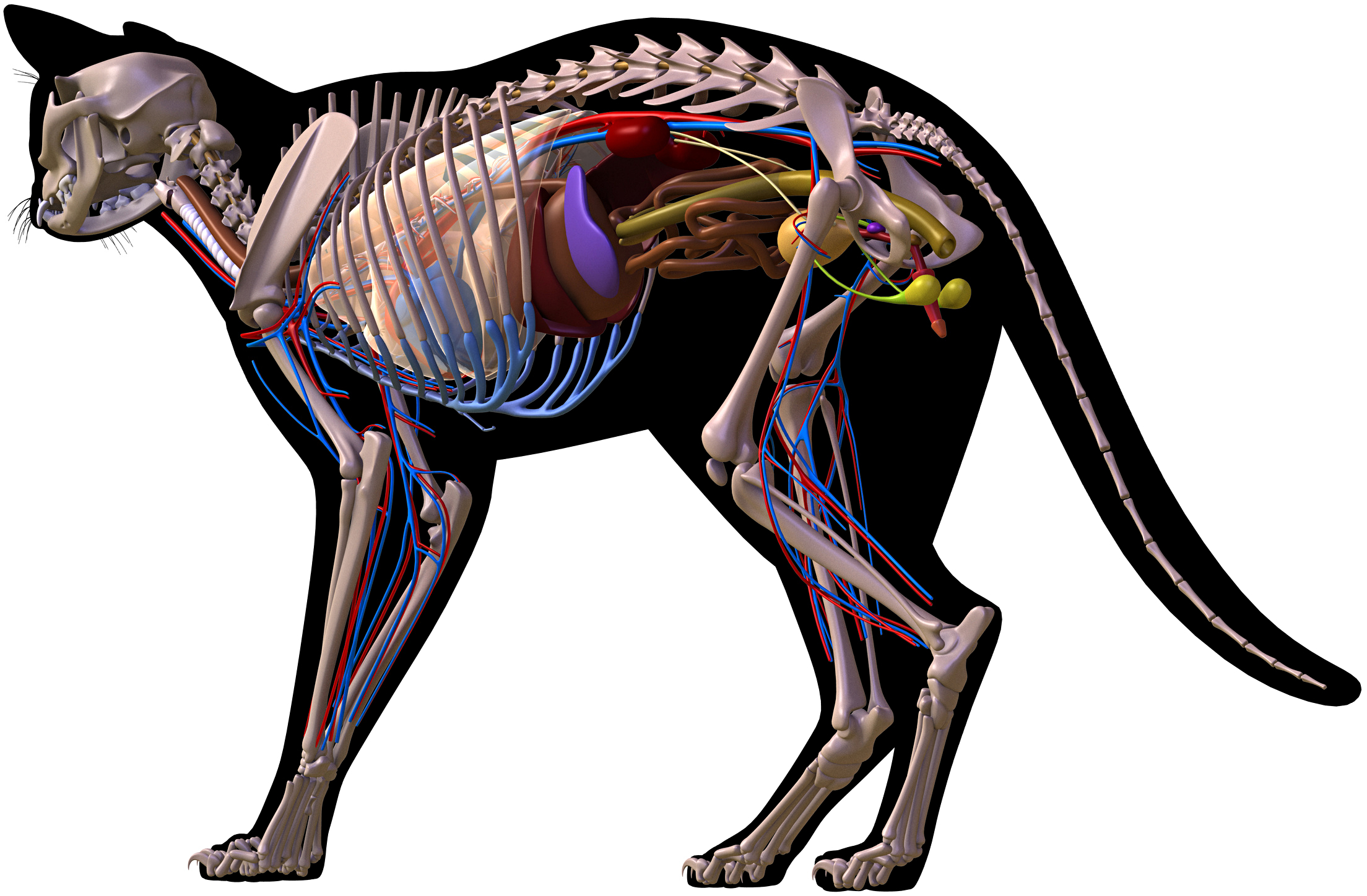
The cat muscle anatomy includes the origin, insertion, and fiber direction of every single muscle from the different regions of the body. Here, I will show you the essential muscles from the face, neck, forelimb, abdomen, and hindlimb. You will also find the description of these muscles from the different regions.
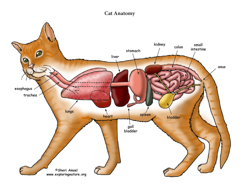
Cat Anatomy (Thoracic and Abdominal Organs)
The aim of the present review is to describe the anatomy of the gastrointestinal tract of the abdomen in cats in combination with the surgical procedures that are performed in each region, highlighting the points of surgical interest.
abdomen internal structure
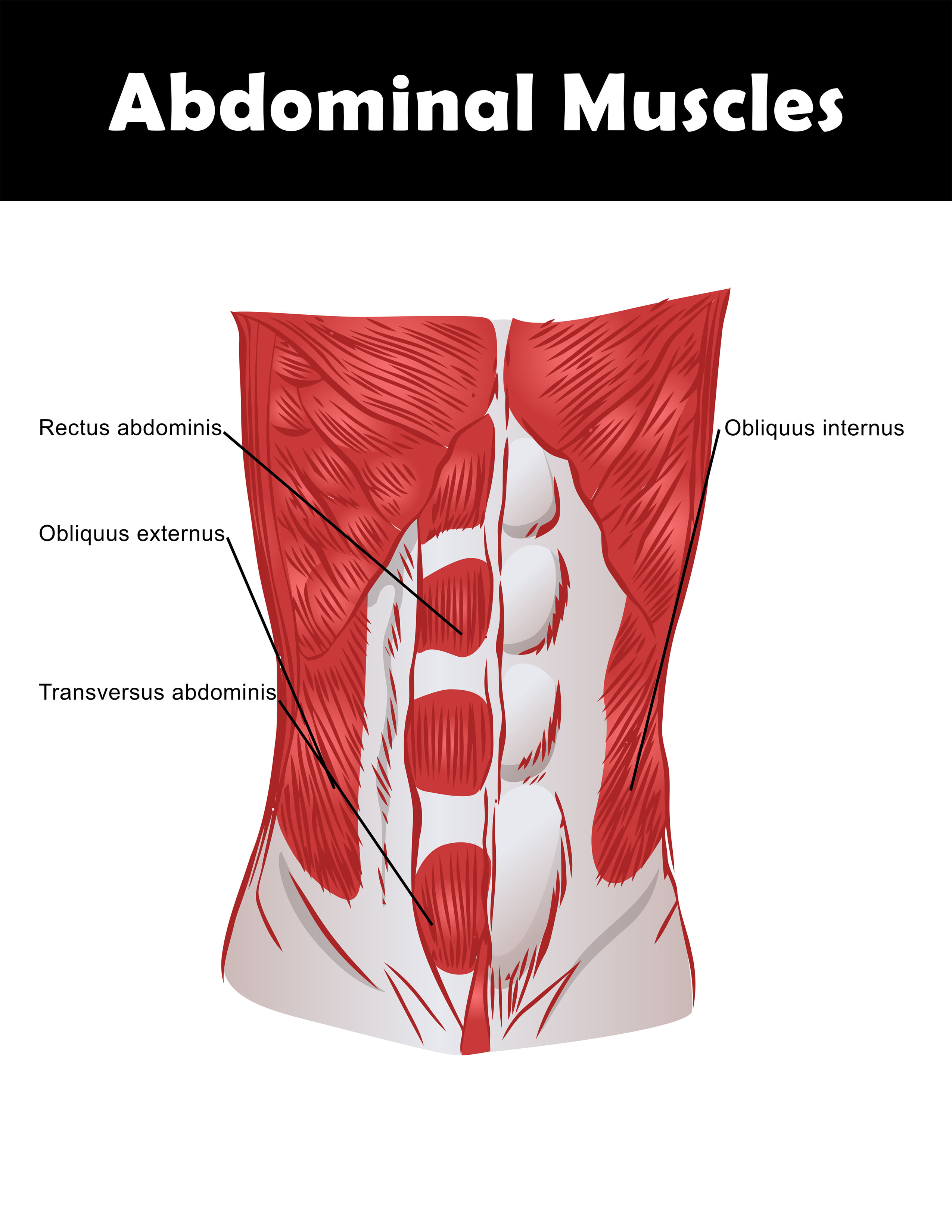
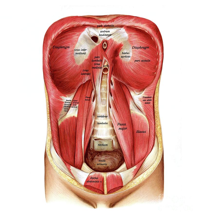
right colic vessels. right common iliac artery. abdominal portion of the ureter. left colic artery. umbilicus. ileocecal valve. left common iliac artery. quadratus lumborum muscle. transversus abdominis muscle.
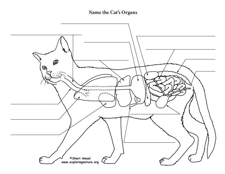
Cat Anatomy (Thoracic and Abdominal Organs)
Quick Overview. 01. Every aspect of your cat's anatomy is fine-tuned for their status as predatory animals. 02. Cats have powerful senses of smell and hearing, making them keenly aware of their environment. 03. Your cat's facial expressions, from whiskers to ears to eyes, can tell you how they're feeling.
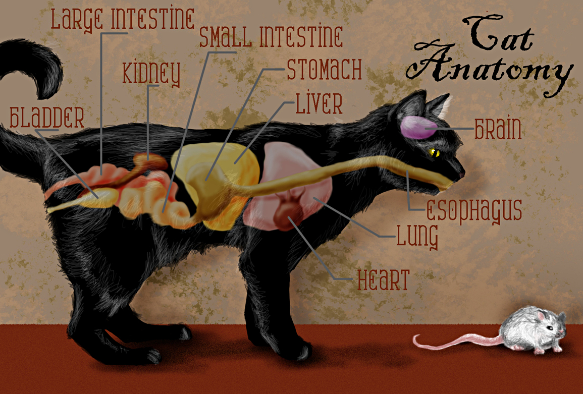
Links to Pictures on the Physiology of Cats
ISSN 2534-5087. This module of vet-Anatomy is a basic atlas of normal imaging of anatomical feline radiology. The 39 sampled x-ray images of healthy cats were performed by Susanne AEB Borofka (PhD - dipl. ECVDI, Utrecht, Netherland). Those images were categorized topographically into six chapters (head, vertebral column, thoracic limb, pelvic.

Cybex Abdominal Primo Fitness
Peritoneal Anatomy 1:53 ; CT Anatomy 21:10 ; Approach 56:00 ; If you want to learn how to read CT scans of the abdomen and pelvis proficiently, this video is an excellent starting point..

Abdominal ultrasound anatomy Small Animal Ultrasonography
Computed tomography ( CT or CAT scan) is one of the most commonly used medical imaging procedures in clinical practice, along with radiography (x-ray) and magnetic resonance imaging (MRI).
Abdominal Anatomy Posterior Posterior Abdomen Abdominal surface
Computed tomography (CT scan or CAT scan) is a noninvasive diagnostic imaging procedure that uses a combination of X-rays and computer technology to produce horizontal, or axial, images (often called slices) of the body. A CT scan shows detailed images of any part of the body, including the bones, muscles, fat, organs, and blood vessels.

Anatomía del gato, Anatomía del perro, Anatomia veterinaria
Cat Anatomy (Thoracic and Abdominal Organs) High Resolution PDF for Printing. Click Here. Link to More Information About This Animal. Click Here. Citing Research References. When you research information you must cite the reference. Citing for websites is different from citing from books, magazines and periodicals. The style of citing shown.

Muscles , 5 Cat Muscle Anatomy Diagram Cat Muscles Ventral Region Key
2021 Ultimate Guide to Cat Anatomy. As the pace of veterinary advancement accelerates, even the most experienced veterinary teams are challenged to keep up with all the changes that impact their practice.. Females undergo a spey or ovariohysterectomy which requires abdominal surgery to remove the uterus and ovaries. Male genitalia. A.
